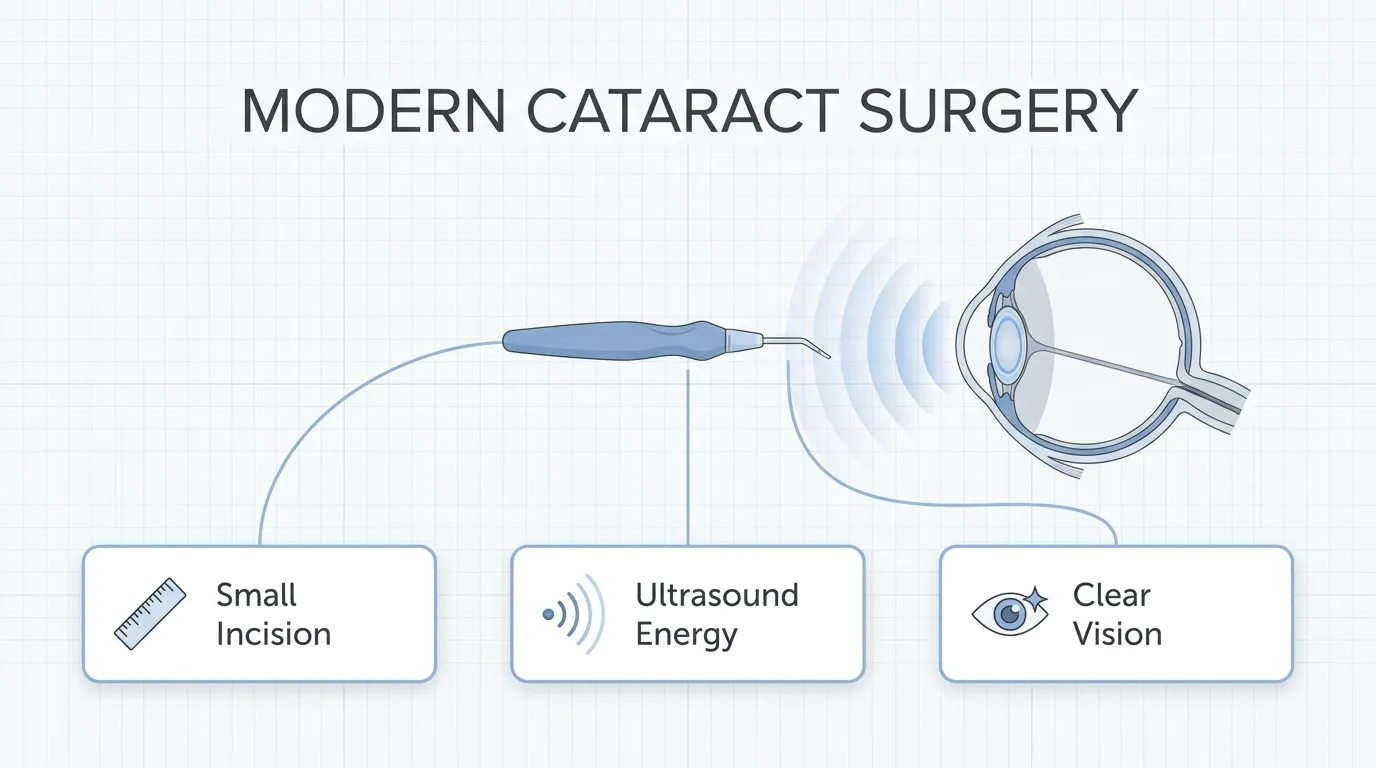
Phacoemulsification is a cataract surgery technique that uses ultrasonic energy to fragment and remove a clouded natural lens through a small incision, typically 2 to 3 mm, before replacing it with an artificial intraocular lens (IOL).
This guide covers how the procedure works and who qualifies, its potential benefits and associated risks, the recovery process and return to daily activities, comparisons with alternative cataract techniques, IOL options available during surgery, and current success rates.
Phacoemulsification operates through a precise sequence of steps, from eye preparation and microincision creation to ultrasound lens fragmentation and IOL implantation. Not every patient is a candidate; eligibility depends on ocular health, systemic conditions, and whether surgery is likely to produce meaningful visual improvement.
The procedure’s small incision may promote faster healing, reduced astigmatism, and a quicker return to normal activities compared to older, larger-incision methods. Many patients notice improved vision within days, though full recovery generally spans four to eight weeks.
Potential complications include posterior capsule opacification, endophthalmitis, corneal edema, cystoid macular edema, and retinal detachment, though incidence rates for serious adverse events have declined over the past two decades.
Patients can choose from monofocal, multifocal, toric, and extended depth of focus IOLs, each addressing different visual needs and lifestyle priorities. Lens selection is one of the most consequential decisions in the entire process.
With millions of procedures performed globally each year, phacoemulsification remains the dominant cataract surgery approach, supported by a well-documented safety profile and consistently strong visual outcomes.
What Does Phacoemulsification Mean in Ophthalmology?
Phacoemulsification means a modern cataract surgery technique that uses ultrasonic energy to emulsify and remove a clouded natural lens through a small incision. The term derives from the Greek word “phakos,” meaning lens, combined with “emulsification,” describing the process of breaking the lens into tiny fragments for aspiration. Charles Kelman first developed this technique in 1967, fundamentally changing how ophthalmologists approach cataract removal.
Unlike older methods requiring large incisions, phacoemulsification operates through an opening of just 2 to 3 mm. A specialized ultrasonic probe, powered by piezoelectric crystals, converts electrical energy into mechanical vibrations at frequencies typically ranging from 27,000 to 60,000 Hz. These high-frequency vibrations fragment the hardened lens material into small pieces, which are then suctioned from the eye. According to StatPearls, phacoemulsification is a modern cataract surgery where a cataractous lens is emulsified through a small incision using an ultrasonic probe.
After the clouded lens is removed, an artificial intraocular lens replaces it, restoring clear vision. The small incision size is one of the procedure’s most significant advantages; it often self-seals without sutures, which can promote faster healing and reduce surgically induced astigmatism. With between 25 and 30 million cataract operations performed globally each year, phacoemulsification remains the dominant surgical approach in modern ophthalmology. For patients exploring whether this procedure fits their needs, understanding how phacoemulsification works step by step offers a clearer picture of what to expect.
How Does Phacoemulsification Work Step by Step?
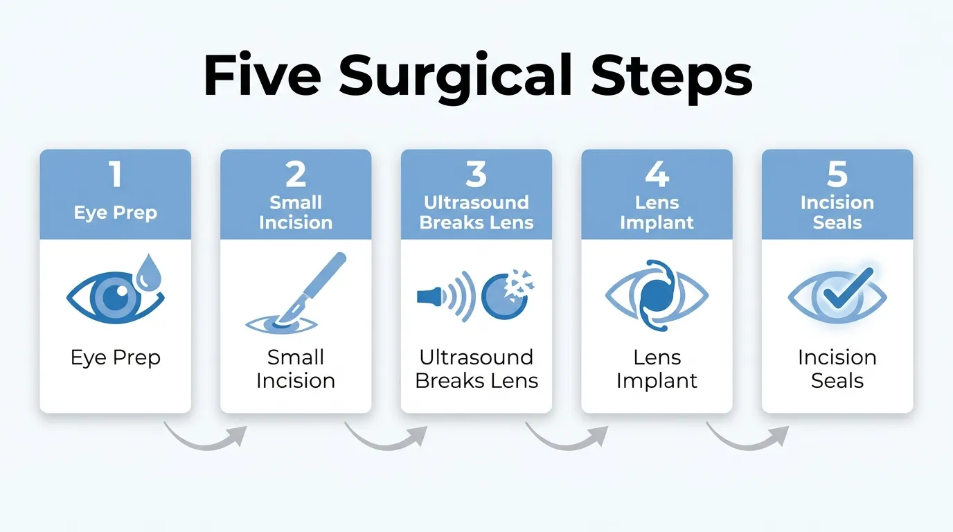
Phacoemulsification works step by step through a sequence of five stages: eye preparation, incision creation, ultrasound lens fragmentation, intraocular lens implantation, and incision sealing.
How Is the Eye Prepared Before Phacoemulsification?
The eye is prepared before phacoemulsification through a series of steps designed to ensure patient comfort and optimal surgical conditions. The ophthalmologist administers topical anesthetic drops to numb the corneal surface, eliminating pain during the procedure. Pupil-dilating drops are then applied to widen the pupil, giving the surgeon a clear path to the cataract behind the iris.
An antiseptic solution, typically povidone-iodine, is used to cleanse the periocular skin and conjunctival surface. This step may help reduce the risk of postoperative infection. A sterile drape and lid speculum are placed to keep the eyelids open and maintain an unobstructed surgical field. Thorough preparation at this stage sets the foundation for each subsequent step in the procedure.
How Does the Surgeon Create the Incision?
The surgeon creates the incision by making a small, self-sealing cut in the peripheral cornea, typically measuring 2 to 3 millimeters in width. This microincision is constructed with a specialized keratome blade at a precise angle, forming a tunnel-like architecture through the corneal layers.
A secondary paracentesis, or side-port incision, is often made to allow insertion of additional instruments. Viscoelastic material is then injected into the anterior chamber to maintain its shape and protect the delicate corneal endothelium during surgery. The small incision size is one of the defining advantages of phacoemulsification, as it typically requires no sutures and promotes faster visual recovery compared to larger-incision techniques.
How Is Ultrasound Used to Break Up the Cataract?
Ultrasound is used to break up the cataract by delivering high-frequency mechanical vibrations through a titanium or steel phaco needle inserted into the lens. According to StatPearls, piezoelectric crystals within the handpiece convert electrical energy into mechanical energy at frequencies typically ranging from 27,000 to 60,000 Hz, fragmenting the hardened lens material into tiny emulsified particles.
The surgeon performs a capsulorhexis, a circular opening in the anterior lens capsule, before accessing the cataract nucleus. Techniques such as divide-and-conquer or phaco chop are used to section the nucleus into manageable quadrants. Simultaneously, an irrigation-aspiration system flushes balanced salt solution into the eye while suctioning out emulsified lens fragments. Careful energy management during this step is critical, as excessive ultrasound can stress the corneal endothelium.
How Is the Intraocular Lens Implanted?
The intraocular lens is implanted by folding a synthetic IOL and inserting it through the same microincision used for cataract removal. The folded lens is loaded into a specialized injector cartridge and guided into the empty capsular bag, where it gently unfolds and centers itself using flexible haptics.
Surgeons select the IOL type and power based on preoperative biometric measurements. According to a 2026 survey published by Review of Ophthalmology, 33% of surgeons chose the Alcon Clareon as their preferred monofocal lens, while 39.7% selected the Alcon Vivity as their top presbyopia-correcting option. IOL selection remains one of the most consequential decisions in the entire procedure, directly shaping each patient’s postoperative visual experience.
How Is the Incision Sealed After Surgery?
The incision is sealed after surgery by hydrating the corneal wound edges with balanced salt solution, causing the tissue to swell slightly and close the self-sealing tunnel without sutures. The surgeon carefully checks the incision for leaks by applying gentle pressure and observing for any fluid escape.
Residual viscoelastic material is aspirated from the anterior chamber to prevent postoperative intraocular pressure spikes. While the self-sealing design provides reliable closure in the vast majority of cases, a study published by the National Center for Biotechnology Information found the overall 10-year incidence of retained lens fragments after phacoemulsification to be approximately 1.47%, underscoring the importance of thorough cortical cleanup before wound closure. Antibiotic and anti-inflammatory drops are applied, and a protective shield is placed over the eye.
With each surgical step complete, the next consideration is determining who qualifies for this procedure.
Who Is a Good Candidate for Phacoemulsification?
A good candidate for phacoemulsification is typically someone whose cataracts cause significant vision impairment that affects daily activities. The subsection below covers situations where this procedure may not be appropriate.
Who May Not Be Eligible for Phacoemulsification?
Patients who may not be eligible for phacoemulsification include those with certain clinical or personal factors that reduce the likelihood of a safe or beneficial outcome. According to StatPearls (NCBI Bookshelf), contraindications for phacoemulsification include multiple systemic comorbidities, patient unwillingness or lack of desire for surgery, good visual acuity with glasses, and situations where surgery will not improve vision or postoperative care is not possible.
Additional factors that may limit eligibility include:
- Uncontrolled systemic conditions, such as diabetes or hypertension, that can increase surgical risk.
- Active ocular infections or inflammation that must be resolved before any intraocular procedure.
- Inability to comply with postoperative care instructions, including prescribed eye drops and follow-up visits.
- Unrealistic expectations about visual outcomes, particularly when underlying retinal or optic nerve disease limits potential improvement.
Your ophthalmologist may recommend alternative approaches or delay surgery until conditions stabilize. Thorough preoperative screening helps identify these factors early, which is why preparation before surgery plays such a critical role.
How Should You Prepare for Phacoemulsification?
You should prepare for phacoemulsification by following your ophthalmologist’s preoperative instructions, which typically cover medication adjustments, eye measurements, and day-of-surgery logistics. Proper preparation helps reduce complications and supports a smoother recovery.
Your eye surgeon may recommend the following steps in the days and weeks before your procedure:
- Complete a comprehensive eye exam. Preoperative testing typically includes ocular biometry to measure axial length, lens thickness, and white-to-white distance, all of which are used to calculate the correct intraocular lens power for your eye.
- Disclose your full medication list. Blood thinners, alpha-blockers for prostate conditions, and certain supplements may need to be paused or adjusted before surgery. Your surgeon will provide specific guidance based on your health profile.
- Begin prescribed eye drops. Many surgeons prescribe antibiotic or anti-inflammatory drops one to three days before the procedure to reduce infection risk and minimize postoperative inflammation.
- Arrange transportation home. Because sedation and pupil dilation affect your ability to drive, you will need someone to take you home after surgery.
- Fast as directed. If intravenous sedation is planned, your surgical team may ask you to stop eating and drinking several hours beforehand.
- Avoid eye makeup and facial lotions on the day of surgery. These products can introduce contaminants near the surgical site.
According to CRSToday, certain ocular biometrics, including axial length, white-to-white distance, and lens thickness, are fundamental parameters for IOL power calculations and preoperative screening before refractive cataract surgery and other refractive laser treatments. This preoperative measurement step is arguably the most consequential part of preparation, since an inaccurate IOL calculation can compromise your visual outcome regardless of how well the surgery itself is performed.
If you have systemic health conditions, such as diabetes or high blood pressure, your ophthalmologist may coordinate with your primary care provider to ensure these are well controlled before proceeding. Patients who cannot commit to postoperative care or follow-up visits may need to address those barriers before scheduling surgery.
Thorough preparation sets the stage for what happens next in the operating room.
What Can You Expect During the Phacoemulsification Procedure?
During the phacoemulsification procedure, you can expect a brief outpatient surgery performed under local anesthesia, typically lasting 15 to 30 minutes per eye.
The experience begins in a preoperative area where the surgical team prepares you and your eye. Once in the operating room, the surgeon works through a small incision while you remain awake but comfortable. Most patients report feeling only mild pressure, not pain, throughout the process.
Your surgeon dilates the pupil with eye drops well before the procedure starts. Topical or local anesthetic drops numb the eye’s surface, eliminating sharp sensation. In some cases, a mild sedative may be administered intravenously to ease anxiety. An eyelid speculum gently holds the eye open, so blinking is not a concern. During surgery, you may see bright lights or shifting colors, which is normal as the microscope illuminates the surgical field.
After the ultrasound probe emulsifies the clouded lens and the new intraocular lens is placed, the surgeon verifies proper positioning. The small incision typically self-seals without sutures. You are then moved to a recovery area for brief monitoring before being discharged the same day, usually within an hour. A protective eye shield is placed over the treated eye to guard against accidental contact.
In clinical practice, patients who understand these steps beforehand tend to feel significantly calmer during surgery. Knowing what each sensation means, from the pressure of the speculum to the bright light of the microscope, removes much of the uncertainty that fuels anxiety.
With a clear picture of what the procedure involves, the next step is understanding the specific benefits phacoemulsification may offer for your vision and recovery.
What Are the Potential Benefits of Phacoemulsification?
The potential benefits of phacoemulsification include improved visual acuity, faster healing from a small incision, reduced dependence on glasses, and shorter procedure time. For those considering broader vision enhancements, various cosmetic eye procedures, such as eyelid surgery, also offer significant aesthetic and functional improvements.

How May Phacoemulsification Improve Visual Acuity?
Phacoemulsification may improve visual acuity by removing the clouded natural lens and replacing it with a clear intraocular lens (IOL) that restores light transmission to the retina. For most patients with visually significant cataracts, this exchange can dramatically sharpen both distance and near vision.
Not every patient, however, is expected to achieve the same degree of improvement. According to StatPearls, contraindications for phacoemulsification include situations where surgery will not improve vision, multiple systemic comorbidities, and cases where postoperative care is not possible. A thorough preoperative evaluation helps the ophthalmologist determine whether the procedure is likely to produce meaningful visual gains for each individual. When candidacy criteria are met, phacoemulsification remains one of the most reliable methods for restoring functional sight compromised by cataract formation.
How Does the Small Incision Promote Faster Healing?
The small incision in phacoemulsification promotes faster healing because it measures only 2 to 3 mm, minimizing disruption to corneal tissue. Less tissue trauma means:
- Reduced postoperative inflammation at the surgical site.
- Lower risk of surgically induced astigmatism.
- Self-sealing wound architecture, often eliminating the need for sutures.
- Quicker stabilization of the corneal surface.
Because the incision is so narrow, the structural integrity of the eye remains largely intact throughout the procedure. This translates to a shorter inflammatory response and a more comfortable recovery compared with larger-incision techniques. In clinical practice, the self-sealing nature of micro-incision wounds is one of the most underappreciated advantages of modern phacoemulsification.
How Can Phacoemulsification Reduce Dependence on Glasses?
Phacoemulsification can reduce dependence on glasses by allowing the surgeon to select an IOL power customized to the patient’s refractive needs. During preoperative planning, precise biometric measurements guide IOL calculations designed to correct preexisting refractive errors, including myopia, hyperopia, and astigmatism.
Depending on the IOL type chosen, patients may experience significantly less reliance on corrective eyewear for daily tasks. Toric IOLs address corneal astigmatism directly, while multifocal and extended depth of focus lenses provide functional vision across multiple distances. Complete spectacle independence is not guaranteed for every patient, yet many find their need for glasses substantially reduced after surgery.
How Does Shorter Procedure Time Help Patients?
Shorter procedure time helps patients by reducing exposure to anesthesia, lowering intraoperative stress, and decreasing the overall physiological burden of surgery. Phacoemulsification typically takes 15 to 30 minutes per eye, making it well suited for outpatient settings.
According to the Cleveland Clinic, while full recovery from cataract surgery typically takes four to eight weeks, many patients notice significant improvement in their vision within a few days following the procedure. The brief operative window also benefits patients with cardiovascular or respiratory conditions who may not tolerate prolonged surgical sessions. For most individuals, the combination of rapid procedure time and early visual recovery makes phacoemulsification a practical, low-disruption intervention.
With its clinical advantages established, understanding the possible risks and complications provides a complete picture before making a surgical decision.
What Are the Possible Risks and Complications?
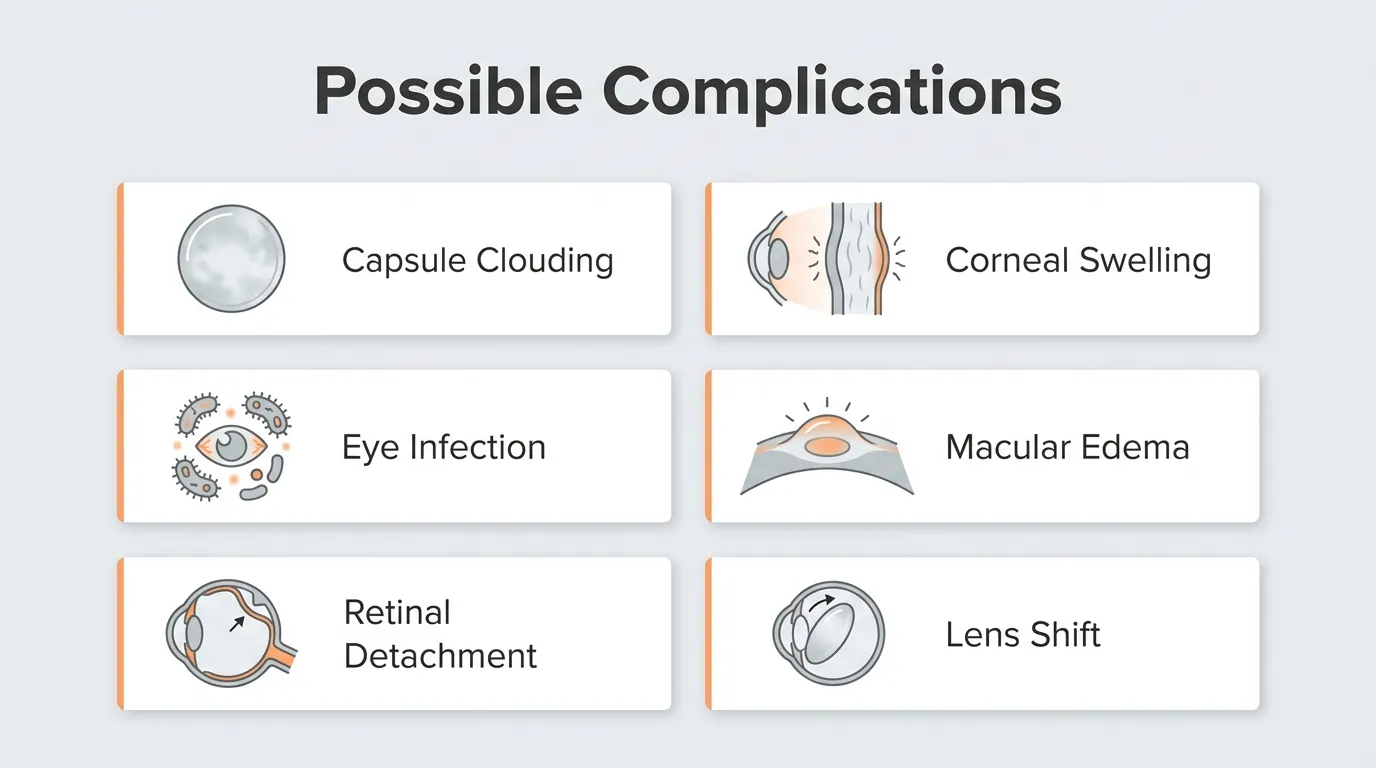
The possible risks and complications of phacoemulsification include posterior capsule opacification, endophthalmitis, retinal detachment, corneal edema, cystoid macular edema, and intraocular lens dislocation.
What Is Posterior Capsule Opacification After Phacoemulsification?
Posterior capsule opacification (PCO) is the most common long-term complication after phacoemulsification. PCO occurs when residual lens epithelial cells proliferate across the posterior capsule, causing blurred vision weeks to months after surgery. A 2026 systematic evaluation published in the International Journal of Surgery reported that the incidence of Nd:YAG posterior capsulotomy, the standard treatment for PCO, was 0.5% at 6 months postoperatively, increasing to 3.7% at 12 months. The Nd:YAG laser capsulotomy procedure is quick, painless, and performed in an outpatient setting. While PCO can be concerning for patients, it is highly treatable and does not diminish the long-term visual gains achieved through the original cataract surgery.
What Is the Risk of Endophthalmitis?
The risk of endophthalmitis after phacoemulsification is low but clinically significant due to its potential to cause severe vision loss. Endophthalmitis is an intraocular infection typically caused by bacteria entering the eye during or shortly after surgery. According to a 2022 report published in the Review of Optometry, the incidence of postprocedural endophthalmitis following intraocular procedures declined significantly from 0.2% in 2000 to 0.05% in 2022. This fourfold reduction reflects advances in sterile technique, prophylactic intracameral antibiotics, and improved surgical protocols. Symptoms such as increasing pain, redness, and decreased vision within days of surgery warrant immediate evaluation. Early detection and treatment with intravitreal antibiotics can preserve functional vision in many cases.
Can Phacoemulsification Cause Retinal Detachment?
Yes, phacoemulsification can cause retinal detachment, though the risk remains relatively uncommon. Retinal detachment occurs when the retina separates from its underlying supportive tissue, potentially leading to permanent vision loss if untreated. Patients with high myopia or prior ocular trauma may face elevated risk. Symptoms to monitor include sudden flashes of light, new floaters, or a shadow spreading across the visual field. Prompt surgical intervention, such as vitrectomy or scleral buckle, can often reattach the retina successfully. For most patients undergoing routine phacoemulsification, this complication is rare, but awareness of warning signs remains essential for early treatment.
What Is the Risk of Corneal Edema?
The risk of corneal edema after phacoemulsification is generally low and typically transient. Corneal edema involves swelling of the corneal tissue caused by endothelial cell damage during ultrasonic lens emulsification. Patients with pre-existing endothelial compromise, such as Fuchs dystrophy, face higher susceptibility. Mild corneal edema often resolves within days to weeks with topical hypertonic saline drops and anti-inflammatory medication. Persistent edema affecting vision may indicate significant endothelial cell loss, which in rare cases requires corneal transplantation. Modern phacoemulsification techniques that minimize ultrasound energy exposure have meaningfully reduced the incidence of clinically significant corneal edema.
Can Cystoid Macular Edema Develop After Surgery?
Yes, cystoid macular edema (CME) can develop after phacoemulsification surgery. CME involves fluid accumulation in the macula, the central retinal area responsible for sharp vision. According to data published in the Expert Review of Ophthalmology (Taylor & Francis Online), pseudophakic CME presented a pooled incidence of 2.41% in specific study populations, higher than the 1.17% incidence observed in the general population. Risk factors include diabetes, uveitis history, and complicated surgical cases. Topical nonsteroidal anti-inflammatory drugs (NSAIDs) and corticosteroids are the standard prophylactic and treatment approach. Most cases of pseudophakic CME resolve with medical therapy, though persistent cases may require intravitreal injections.
What Is the Risk of Intraocular Lens Dislocation?
The risk of intraocular lens (IOL) dislocation after phacoemulsification is low. IOL dislocation occurs when the implanted lens shifts from its intended position within the capsular bag, potentially causing visual disturbances such as diplopia, glare, or refractive changes. Zonular weakness, pseudoexfoliation syndrome, and prior ocular trauma are recognized predisposing factors. Late dislocation, occurring years after surgery, is more common than early displacement. Surgical repositioning or IOL exchange can restore proper lens alignment and visual function when dislocation occurs. This complication, while uncommon, reinforces the importance of long-term follow-up after cataract surgery.
Understanding these potential complications helps patients recognize warning signs early and seek timely care from their ophthalmologist.
What Does Recovery After Phacoemulsification Look Like?
Recovery after phacoemulsification typically takes four to eight weeks for full healing, though vision often improves within the first few days. The subsections below cover the first week, weeks two through four, and the timeline for returning to normal activities.
What Happens During the First Week of Recovery?
The first week of recovery after phacoemulsification involves the most careful postoperative management. Your ophthalmologist may prescribe antibiotic and anti-inflammatory eye drops to reduce infection risk and control swelling. According to the Cleveland Clinic, many patients notice a significant improvement in their vision within a few days following the procedure, even as the eye continues to heal.
During this initial period, mild discomfort, light sensitivity, and slight blurriness are common. A protective eye shield is typically worn at night to prevent accidental rubbing or pressure. Patients are generally advised to avoid:
- Touching or pressing on the operated eye
- Heavy lifting or strenuous physical activity
- Getting water directly in the eye while bathing
- Dusty or smoky environments that could irritate healing tissue
In most cases, a follow-up appointment occurs within the first 24 to 48 hours so the surgeon can check intraocular pressure and confirm that the incision site is sealing properly.
What Should You Expect During Weeks Two Through Four?
During weeks two through four, you should expect gradual visual stabilization as the eye continues healing. Residual blurriness and minor fluctuations in vision may persist, but these typically diminish steadily throughout this period.
Anti-inflammatory drops often continue during this phase, while antibiotic drops may be tapered based on your surgeon’s assessment. The small, self-sealing incision used in phacoemulsification promotes faster tissue recovery compared to older, larger-incision techniques, which is why most patients feel comfortable resuming light daily routines relatively early.
Some sensitivity to bright light can linger into the fifth or sixth week. Wearing sunglasses outdoors helps protect the eye during this transitional phase. Your ophthalmologist may schedule one or two additional follow-up visits to monitor healing progress and refine any updated eyeglass prescription, since refractive measurements taken too early may not reflect your final visual outcome.
When Can You Return to Normal Activities?
You can return to most normal activities within a few days to several weeks after phacoemulsification, depending on the activity’s physical demands. Light tasks, such as reading, watching television, and gentle walking, are generally safe within the first few days.
A practical timeline for resuming common activities includes:
- Light desk work and reading: within several days
- Driving: once your surgeon confirms adequate visual acuity, often within one week
- Light exercise such as walking: within one week
- Swimming and hot tubs: typically after four weeks to reduce infection risk
- Heavy lifting and contact sports: once your surgeon confirms adequate healing
Full recovery from phacoemulsification generally spans four to eight weeks, though individual healing rates vary based on age, overall health, and whether any complications arise. Your ophthalmologist’s guidance should always take priority over general timelines, since each patient’s healing trajectory is unique. With a clear recovery plan in place, the next step is understanding how phacoemulsification compares to alternative cataract techniques.
How Does Phacoemulsification Compare to Other Cataract Techniques?
Phacoemulsification compares to other cataract techniques through differences in incision size, energy delivery, and recovery timelines. The subsections below examine how it differs from ECCE and femtosecond laser-assisted surgery.
How Does Phacoemulsification Compare to ECCE?
Phacoemulsification compares to ECCE (extracapsular cataract extraction) primarily in incision size and recovery speed. ECCE requires a large incision, typically 10 to 12 mm, to remove the lens nucleus in one piece. Phacoemulsification uses an ultrasonic probe through an incision of roughly 2 to 3 mm, fragmenting the lens before aspiration.
This smaller incision generally promotes faster wound healing, reduces surgically induced astigmatism, and allows most patients to resume normal activities sooner. ECCE may still be preferred in certain clinical scenarios, such as very dense or mature cataracts where ultrasonic emulsification would require excessive energy. For the majority of routine cataract cases, however, phacoemulsification offers a more controlled surgical environment with quicker visual rehabilitation. Surgeons typically reserve ECCE for situations where phacoemulsification poses a higher risk of complications.
How Does Phacoemulsification Compare to Femtosecond Laser-Assisted Surgery?
Phacoemulsification compares to femtosecond laser-assisted surgery (FLACS) in how each technique fragments the cataractous lens. Conventional phacoemulsification relies entirely on ultrasonic energy delivered through a handheld probe. FLACS adds a femtosecond laser to pre-treat the lens, softening and fragmenting it before the ultrasonic probe completes the removal.
According to a systematic review and meta-analysis published by Scientific Research Publishing, FLACS was associated with a reduction in effective phacoemulsification time and cumulative dissipated energy compared with conventional phacoemulsification in patients with dense cataracts. Less ultrasonic energy may translate to reduced stress on corneal endothelial cells, which is particularly relevant for patients with compromised corneal health.
Despite these advantages, FLACS involves higher procedural costs and additional equipment. For most standard cataract cases, conventional phacoemulsification delivers excellent outcomes without the added expense. FLACS tends to offer the greatest clinical benefit in complex or dense cataracts where minimizing ultrasound exposure matters most. Understanding these trade-offs can help when exploring the types of intraocular lenses available for implantation.
What Types of Intraocular Lenses Can Be Used?
The types of intraocular lenses that can be used during phacoemulsification include monofocal, multifocal, toric, and extended depth of focus designs. Each IOL type addresses different visual needs.
Monofocal IOLs
Monofocal IOLs are intraocular lenses designed to provide clear vision at a single focal distance, typically set for distance viewing. Most patients who receive a monofocal lens still need reading glasses for near tasks. These lenses remain the most commonly implanted IOL worldwide due to their reliable optical quality and predictable outcomes. According to a 2026 Review of Ophthalmology survey of IOL preferences, 33% of surgeons chose the Alcon Clareon as their preferred monofocal lens. For patients who prioritize consistent distance clarity without the potential visual trade-offs of advanced designs, monofocal IOLs often represent the most straightforward choice.
Multifocal IOLs
Multifocal IOLs are intraocular lenses that use concentric rings or zones to split incoming light, providing focus at multiple distances simultaneously. This design can reduce or eliminate the need for glasses after cataract surgery for both near and far tasks. Some patients may notice halos or glare around lights at night, particularly during the initial adaptation period. These visual phenomena tend to diminish as the brain adjusts over several weeks. Multifocal lenses work best for patients motivated to minimize spectacle dependence and who understand the optical trade-offs involved.
Toric IOLs
Toric IOLs are intraocular lenses specifically engineered to correct pre-existing corneal astigmatism during cataract surgery. Unlike standard spherical lenses, toric designs incorporate different optical powers along specific meridians to neutralize the irregular corneal curvature that causes blurred vision. Precise rotational alignment during implantation is critical, because even small degrees of misalignment can reduce astigmatic correction. Patients with moderate to high corneal astigmatism benefit most from toric IOLs, as these lenses address both the cataract and the refractive error in a single procedure.
Extended Depth of Focus IOLs
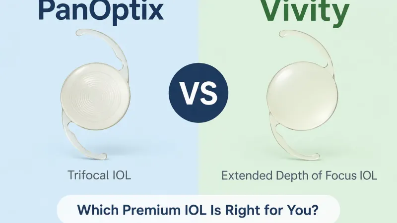
Extended depth of focus IOLs are intraocular lenses that elongate a single focal point rather than splitting light into multiple distinct zones. This design provides a continuous range of clear vision from distance through intermediate tasks, such as computer work, while producing fewer halos and glare than traditional multifocal lenses. The 2026 Review of Ophthalmology survey found that 39.7% of surgeons selected the Alcon Vivity as their top presbyopia-correcting IOL, reflecting growing clinical confidence in EDOF technology. Near vision for fine print may still require reading glasses in some cases, but the smoother visual transition makes EDOF lenses an increasingly popular option for patients seeking functional range with minimal optical disturbances.
With the right lens selected, understanding long-term surgical success rates helps set realistic expectations.
How Successful Is Phacoemulsification in 2026?
Phacoemulsification in 2026 is highly successful, with low complication rates and strong visual outcomes supported by decades of refinement. Between 25 and 30 million cataract operations are performed globally each year, according to a 2026 World Health Organization report, with surgical coverage increasing by approximately 15% over the past two decades despite rising demand from aging populations. This volume reflects the procedure’s established safety profile and widespread clinical confidence.
Complication rates continue to decline. Endophthalmitis incidence dropped from 0.2% in 2000 to 0.05% by 2022. Posterior capsule opacification requiring Nd:YAG capsulotomy occurs in just 0.5% of patients at six months, rising to 3.7% at twelve months. Retained lens fragment incidence remains low at approximately 1.47% over a ten-year period. Pseudophakic cystoid macular edema presents a pooled incidence of 2.41% in specific study populations.
Most patients notice significant visual improvement within days, though full recovery typically takes four to eight weeks. Few surgical procedures in any medical specialty can match this combination of high volume, low complication incidence, and rapid functional recovery. With trusted cataract surgery resources, patients can explore what these outcomes mean for their individual situation.
How Can Trusted Cataract Surgery Resources Help You Decide?
Trusted cataract surgery resources can help you decide by providing evidence-based information that clarifies your procedure options, candidacy factors, and expected outcomes. The following subsections cover how Eye Surgery Today supports your research and the key takeaways from this guide. For parents navigating the unique challenges of childhood cataracts, specific support groups for congenital cataracts offer invaluable emotional and practical assistance.
Can Eye Surgery Today Help You Understand Your Phacoemulsification Options?
Yes, Eye Surgery Today can help you understand your cataract options through its educational platform. Founded by nationally recognized ophthalmology key opinion leaders, Eye Surgery Today translates complex surgical concepts into clear, accessible language designed for patients at every stage of their decision-making process.
Eye Surgery Today provides resources covering critical preoperative considerations. Thorough screening before refractive cataract surgery involves evaluating ocular biometrics and corneal conditions, as highlighted by Cataract & Refractive Surgery Today. Eye Surgery Today breaks down these clinical factors so patients can approach consultations with informed questions about lens selection, surgical technique, and recovery expectations.
From understanding how ultrasonic energy fragments the cataract to comparing IOL categories, Eye Surgery Today offers guides built on evidence-based standards. This level of transparency helps patients move from uncertainty to confidence without relying on oversimplified or biased sources.
What Are the Key Takeaways About Phacoemulsification?
The key takeaways about phacoemulsification are that it remains the global standard for cataract removal, offering small-incision precision, rapid visual recovery, and a well-documented safety profile.
- Phacoemulsification uses ultrasonic energy to fragment and remove the clouded lens through an incision typically measuring 2 to 3 mm.
- Most patients may notice significant vision improvement within days, though full recovery can take four to eight weeks.
- Serious complications, such as endophthalmitis and retinal detachment, are rare and continue to decline with advancing surgical techniques.
- IOL selection directly shapes post-surgical visual outcomes, making preoperative screening and informed discussion with your ophthalmologist essential.
- Not every patient qualifies; candidacy depends on ocular health, systemic conditions, and realistic expectations for visual improvement.
Choosing phacoemulsification is ultimately a collaborative decision between patient and surgeon. The strongest outcomes begin with thorough education, and platforms like Eye Surgery Today exist to ensure that the knowledge gap never stands between patients and parents and clear vision.