Cataract surgery is a lens replacement procedure that removes the eye’s clouded natural lens and replaces it with a clear artificial intraocular lens (IOL). It does not directly treat floaters, which originate in the vitreous gel, a separate structure the surgery leaves untouched.
This guide covers what floaters are and why they appear, how cataract surgery interacts with them, dedicated floater treatments, risk factors for post-surgical floaters, and how to prepare for informed conversations with your surgeon.
Floaters are small shapes (spots, threads, cobwebs) formed by collagen clumps or cellular debris suspended in the vitreous gel. They are extremely common in adults, and conditions like ageing, myopia, and posterior vitreous detachment (PVD) may accelerate their development.
Cataract surgery operates exclusively on the lens in the anterior segment of the eye. Because the vitreous gel in the posterior segment remains intact, pre-existing floaters persist after the procedure. A clearer IOL may also allow more light into the eye, which can make previously masked floaters appear more prominent. In some cases, surgical disturbance to surrounding structures may contribute to new floater onset.
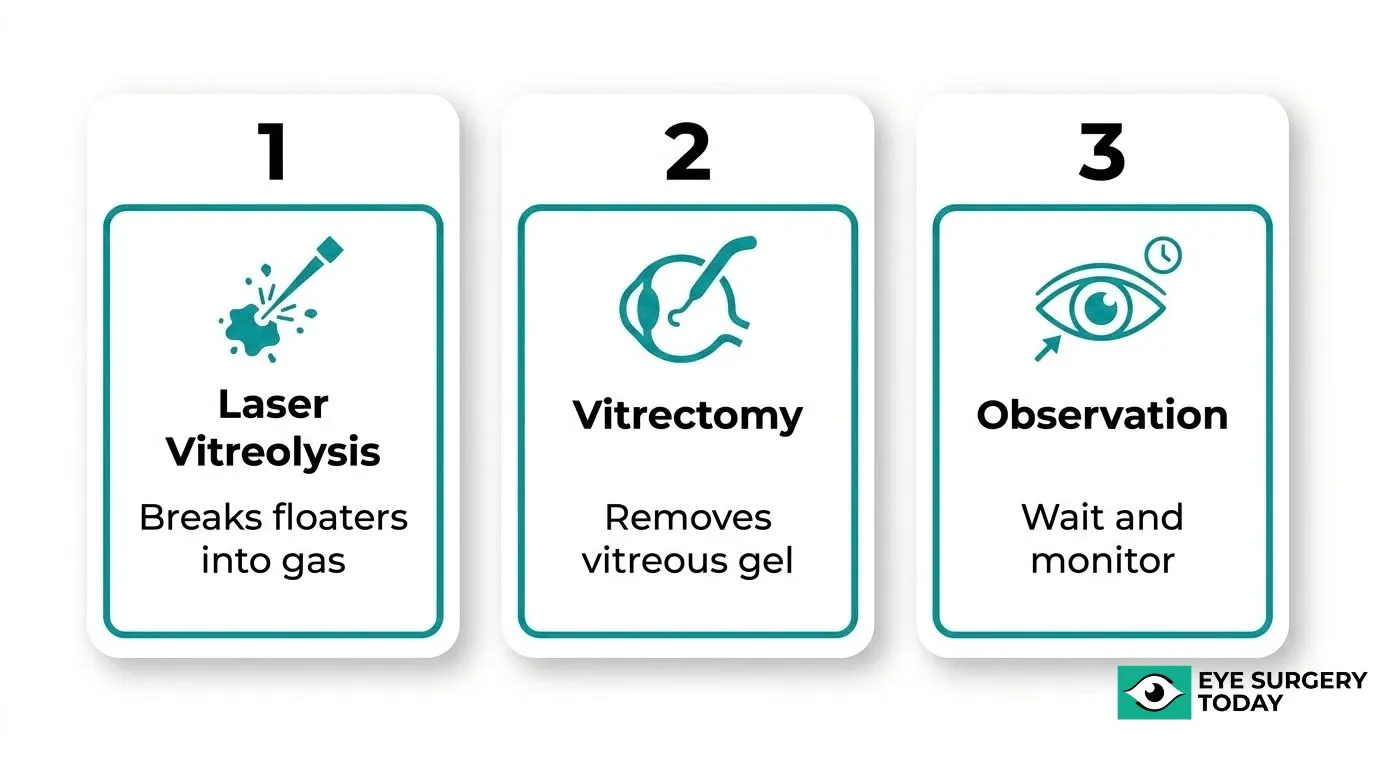
For patients whose floaters significantly affect daily vision, separate interventions exist. Laser vitreolysis uses a YAG laser to break floaters into gas, while vitrectomy surgically removes the vitreous gel entirely. Observation remains a reasonable first approach, as floaters often become less noticeable over time.
Older adults, highly myopic patients, and those with a history of PVD may carry elevated floater risk after cataract surgery. Discussing your floater history, vitreous status, and specific concerns with your surgeon before the procedure helps ensure realistic expectations and a personalized care plan.
What Are Eye Floaters?
Eye floaters are small visual disturbances that drift across your field of vision, originating inside the eye itself. The following sections cover what floaters look like, what causes them, and when they may signal a more serious problem.

What Do Floaters Actually Look Like?
Floaters are small dark shapes that float across your vision. According to the National Eye Institute, they can look like spots, threads, squiggly lines, or even little cobwebs.
These shapes move when your eye moves and tend to drift slowly when your gaze stops. They are most noticeable when looking at a plain, bright background such as a clear sky or white wall.
What Causes Floaters to Appear in Your Vision?
Floaters appear when cells, clumps of collagen proteins, or debris are present within the vitreous gel, which fills the back of the eye. As this material casts shadows on the retina, the brain perceives them as floating shapes drifting across vision, according to Johns Hopkins Medicine.
Several underlying conditions may trigger floaters, including:
- Ageing and natural vitreous shrinkage
- Short-sightedness (myopia)
- Uveitis or eye infection
- Diabetes
- Cataract surgery
Are Floaters a Sign of Something Serious?
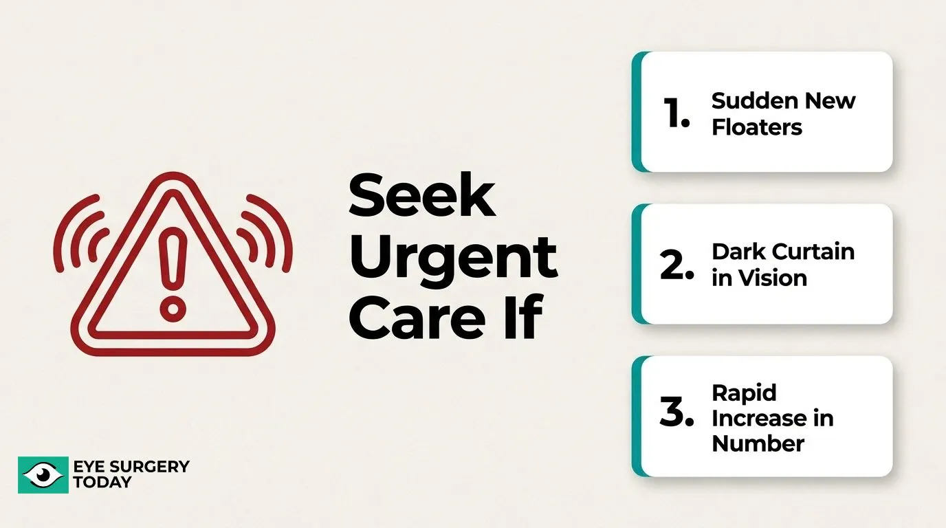
Floaters are usually harmless, but sudden changes in their appearance may indicate a serious underlying condition requiring urgent evaluation. The NHS advises seeking urgent care if you suddenly develop new floaters or flashes, notice a rapid increase in their number, or see a dark curtain or shadow moving across your vision, particularly after eye surgery or injury.
These symptoms may point to a posterior vitreous detachment, retinal tear, or retinal detachment, all of which require prompt assessment to protect vision.
What Happens to Floaters During Cataract Surgery?
Cataract surgery targets the eye’s lens, not the vitreous gel where floaters originate. According to the American Academy of Ophthalmology, during cataract surgery the cloudy natural lens is removed and replaced with a clear artificial intraocular lens (IOL), using a phacoemulsification technique that breaks up the lens with ultrasound and suctions it out. The sections below explain whether this process eliminates floaters, worsens them, or makes them more visible.
Does Removing the Lens Eliminate Floaters?
Removing the lens does not eliminate floaters. Floaters reside in the vitreous humor, the gel-like substance filling the back of the eye, which remains entirely untouched during standard cataract surgery. Because phacoemulsification only addresses the lens, any debris or collagen clumps already present in the vitreous stay in place after the procedure. Patients who enter surgery expecting floater relief are likely to be disappointed, as the two structures are anatomically separate.
Can Cataract Surgery Make Floaters Worse?
Yes, cataract surgery can make floaters worse in some patients. The Macular Society identifies cataract surgery as a recognized cause of new floater onset, alongside inflammation, infection, myopia, diabetes, and ageing. Surgical disturbance to the eye may trigger vitreous changes that produce new floaters postoperatively. This is an important risk for patients to discuss with their surgeon before consenting to the procedure.
Can Cataract Surgery Make Floaters More Noticeable?
Cataract surgery can make floaters more noticeable even when it does not create new ones. Before surgery, a cloudy lens scatters and dims incoming light, which can mask floaters in the visual field. Once a clear IOL replaces the opaque lens, significantly more light enters the eye, making pre-existing vitreous debris suddenly visible and more distracting. Patients should be counseled that improved optical clarity is a feature of successful surgery, but it can bring floaters into sharper perceptual focus.
Why Does Cataract Surgery Not Fix Floaters?
Cataract surgery does not fix floaters because the procedure targets the eye’s lens, while floaters originate in the vitreous gel, a completely separate structure. The following sections explain where floaters come from and why the vitreous remains untouched during lens removal.
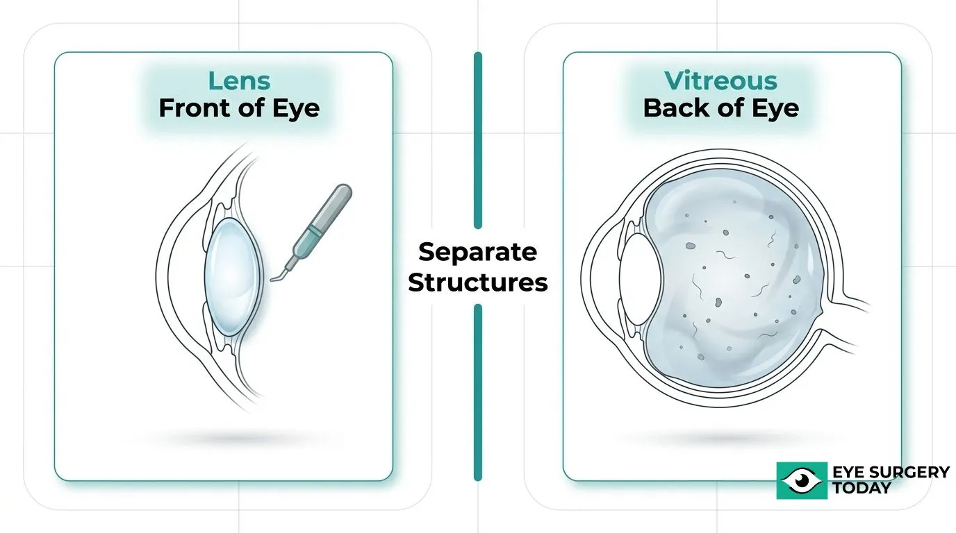
Where Do Most Floaters Actually Come From?
Most floaters come from the vitreous gel that fills the back of the eye, not from the lens that cataract surgery removes. As this gel ages and shrinks, it can pull away from the retina in a process called posterior vitreous detachment (PVD). According to the Australian Family Physician (Royal Australian College of General Practitioners), PVD is the most common cause of acute-onset flashes and floaters, present in nearly 66% of patients over 70. Because the lens and the vitreous are anatomically distinct, removing the lens leaves the vitreous and its debris completely intact.
Why Does the Vitreous Gel Stay Untouched During Cataract Surgery?
The vitreous gel stays untouched during cataract surgery because the procedure is confined to the anterior segment of the eye, where the lens sits, and does not extend to the posterior segment, where the vitreous resides. According to a Johns Hopkins University study published in Experimental Eye Research, while lens cortical fiber debris can leak into the vitreous during cataract extraction, the full extent of vitreous alteration after surgery remains unknown. A separate study in the Journal of Cataract & Refractive Surgery found that in 41.4% of eyes, the vitreous body remained fully attached to the retina one year after surgery. When floaters must be addressed directly, a vitrectomy is required: as the University of California, Irvine Gavin Herbert Eye Institute notes, this procedure removes the vitreous and its floaters, replacing them with a saline solution.
Will Floaters Go Away on Their Own After Cataract Surgery?
Floaters after cataract surgery may resolve on their own, but the outcome depends on their cause and severity. The following sections cover natural resolution timelines and warning signs that require urgent evaluation.
Do New Floaters After Cataract Surgery Disappear Over Time?
New floaters after cataract surgery can disappear over time, but this is not guaranteed for every patient. According to Harvard Health Publishing, Dr. Jeffrey Heier, an ophthalmologist and Harvard Medical School instructor, states that “in many cases, floaters become less noticeable or more tolerable over time, and can even disappear entirely.” A study published in the International Journal of Medical Sciences found that 12.5% of eyes in one surgical group experienced new-onset floater symptoms on the day after cataract surgery, compared to 5.13% and 5.83% in other groups. For many patients, the brain gradually learns to ignore floaters, making them functionally less disruptive even when they do not fully disappear. Persistent or worsening floaters warrant professional evaluation rather than watchful waiting.
When Should You Be Concerned About Floaters Post-Surgery?
You should be concerned about floaters post-surgery when they appear suddenly, increase rapidly, or accompany other visual symptoms. The NHS advises seeking urgent care if floaters or flashes appear for the first time, if the number suddenly increases, if a dark curtain or shadow moves across your vision, or if floaters begin after eye surgery or injury. These symptoms may indicate a retinal tear or detachment, which requires immediate assessment. A PubMed study found that 7.39% of patients with symptomatic posterior vitreous detachment developed delayed retinal tears by 6.24 years after diagnosis, with many occurring well beyond the typical 6-week follow-up window. Prompt evaluation after any sudden change in floaters can be critical for protecting long-term vision.
What Are the Actual Treatments for Floaters?
The actual treatments for floaters are laser vitreolysis, vitrectomy, and watchful waiting. Each option carries different eligibility criteria, risk profiles, and expected outcomes, covered in the H3s below.
What Is Laser Vitreolysis and Does It Work?
Laser vitreolysis is a non-invasive procedure that uses a YAG laser to break down vitreous floaters inside the eye. According to EyeWiki (American Academy of Ophthalmology), the mechanism involves “plasma formation” and “optical breakdown,” where the laser performs photoconversion of the solid floater into a gas through photochemical, thermal, thermoacoustic, and electromagnetic optical field effects.
In terms of efficacy, a study published in The Open Ophthalmology Journal reported a mean improvement in subjective visual symptoms of 71% among treated patients, with no complications detected during follow-up. Safety data from the National Institute for Health and Care Excellence (NICE) confirmed no retinal detachment, retinal tear, uveitis, cystoid macular oedema, or macular holes were identified.
For patients with symptomatic floaters who are not surgical candidates, laser vitreolysis may offer a meaningful improvement in quality of vision with a well-documented safety profile.
What Is Vitrectomy and When Is It Recommended?
Vitrectomy is a surgical procedure that removes the vitreous gel from the eye, along with the floaters it contains, and replaces it with a saline solution. According to the University of California, Irvine Gavin Herbert Eye Institute, vitrectomy may be recommended when floaters begin to drastically affect vision.
While many patients can live with floater symptoms, Calderdale and Huddersfield NHS Foundation Trust notes that some patients experience persistent, disabling floaters that cause genuine visual disability. Recovery typically requires two to four weeks before normal activities can resume, according to Alberta Health Services.
Vitrectomy is generally reserved for severe cases where floaters significantly impair daily function, given that it carries greater surgical risk than laser vitreolysis.
Are There Non-Surgical Options for Managing Floaters?
The non-surgical options for managing floaters are observation and lifestyle adaptation. For many patients, watchful waiting is the first-line approach, as floaters frequently become less noticeable as the brain learns to filter them over time.
According to Harvard Health Publishing, Dr. Jeffrey Heier, an ophthalmologist and Harvard Medical School instructor, notes that “in many cases, floaters become less noticeable or more tolerable over time, and can even disappear entirely.” Importantly, a Cochrane review confirmed there are currently no randomized controlled trials comparing Nd:YAG laser vitreolysis with pars plana vitrectomy, meaning treatment decisions must still rely on clinical judgment and individual patient circumstances. For patients with mild or recently onset floaters, a period of observation is a reasonable and evidence-consistent starting point before pursuing intervention.
Who Is Most Likely to Have Floaters After Cataract Surgery?
Certain patients carry a meaningfully higher risk of floaters after cataract surgery, particularly those who are older, highly myopic, or have a prior history of vitreous detachment. The following H3 sections examine each of these risk profiles in detail.
Are Older Patients More Prone to Floaters After Cataract Surgery?
Older patients are more prone to floaters after cataract surgery, primarily because age accelerates vitreous changes and increases the likelihood of pre-existing posterior vitreous detachment. A large-scale analysis published in Ophthalmology Science, covering over 3.1 million eyes, found that pre-existing PVD was associated with increased odds of retinal detachment post-surgery (odds ratio 1.24; 95% CI, 1.15–1.34). Lattice degeneration, hypermature cataracts, and complex surgical procedures compounded this risk further. Since floaters frequently accompany vitreous traction and PVD, older patients undergoing cataract surgery face a compounding vulnerability that younger patients typically do not.
Do Patients With Myopia Have a Higher Risk of Floaters?
Patients with myopia do have a higher risk of floaters after cataract surgery, largely because longer axial length accelerates vitreous liquefaction and PVD. A study published in Scientific Reports enrolling 170 patients with high myopia (axial length ≥ 26.0 mm) found that increased axial length was associated with more advanced PVD stages, with an odds ratio of 5.16 (95% CI 1.34–19.80) comparing the highest to lowest axial length groups. Research from Washington University School of Medicine further confirmed that among 371 eyes undergoing cataract surgery after prior retinal repair, 4.6% developed a new tear or detachment, with myopia identified as a key risk factor. While most floaters remain benign, clinicians should consider myopia a meaningful risk indicator during pre-surgical planning.
Does a History of Vitreous Detachment Affect Floater Risk?
A history of vitreous detachment does affect floater risk after cataract surgery, as PVD is both a source of existing floaters and a marker of continued vitreous instability. According to the Cleveland Clinic, an estimated 66% of people between the ages of 66 and 86 will develop PVD, making it one of the most prevalent pre-existing conditions in the cataract surgery population. As defined by the University of Iowa EyeRounds, PVD involves separation of the posterior hyaloid face from the neurosensory retina, which may lead to retinal tears and, in more serious cases, retinal detachment. Patients with a documented PVD history should discuss their floater burden and vitreous status with their surgeon well before any lens procedure.
What Should You Tell Your Surgeon About Your Floaters?
Knowing what to share with your surgeon helps ensure safer surgical planning and more realistic expectations. The H3s below cover when to raise floaters and which questions to ask.
When Should You Mention Floaters Before Cataract Surgery?
You should mention floaters before cataract surgery at your very first preoperative consultation. Disclosing your floater history allows your surgeon to assess whether any underlying vitreous or retinal conditions, such as posterior vitreous detachment or retinal tears, may affect surgical risk or postoperative outcomes.
Be sure to share the following information:
- How long you have had floaters and whether they appeared suddenly.
- Whether floaters are accompanied by flashes of light.
- Any history of retinal tears, detachment, or prior eye surgery.
- Whether you have high myopia or a known diagnosis of PVD.
- Any recent increase in the number or size of floaters.
According to the Royal College of Ophthalmologists, the consent process requires that your surgeon discuss the procedure’s likely benefits and risks, along with any particular concerns you raise, giving you the opportunity to ask questions. Bringing floater concerns to that conversation ensures your specific risk profile is documented before surgery proceeds.
What Questions Should You Ask Your Surgeon About Floaters?
The questions you should ask your surgeon about floaters are ones that clarify whether your floaters signal a retinal risk, how surgery may affect them, and what monitoring will follow.
Consider asking:
- Will my existing floaters become more noticeable after cataract surgery?
- Could my floaters indicate posterior vitreous detachment or a retinal tear that needs to be addressed before surgery?
- Am I at higher risk given my age, myopia level, or vitreous history?
- What symptoms after surgery should prompt me to seek urgent care?
- If floaters remain bothersome after surgery, what treatment options are available, such as laser vitreolysis or vitrectomy?
- How often will my retina be monitored during the postoperative period?
Patients who arrive at consultation with specific questions receive more targeted risk guidance. Floaters are a window into vitreous health, and surgeons who understand your full history are better positioned to individualize your care plan.
How Can Patients Make Confident Decisions About Cataract Surgery?
Patients can make confident decisions about cataract surgery by accessing surgeon-reviewed educational resources and understanding the key facts about floaters before and after the procedure. The following H3s cover how credible education supports evaluation and what the most important takeaways are.
Can Surgeon-Reviewed Educational Resources Help You Evaluate Your Options?
Yes, surgeon-reviewed educational resources can help you evaluate your options by providing clinically accurate, unbiased information about what cataract surgery does and does not address. Understanding that the procedure targets the lens while leaving the vitreous gel untouched, for example, helps patients set realistic expectations about floaters.
According to a review published in the Journal of Evidence-Based Medicine, targeted educational interventions significantly increased educational efficacy in 69% of studies, with the most consistent improvements seen in patient understanding of cataract surgery and postoperative care. Well-informed patients ask better questions, recognize warning signs earlier, and are less likely to be caught off guard by outcomes like new or persistent floaters.
Eye Surgery Today provides surgeon-reviewed resources designed to bridge exactly this knowledge gap, translating clinical evidence into clear, accessible guidance for patients considering surgery.
What Are the Key Takeaways About Whether Cataract Surgery Fixes Floaters?
The key takeaways about whether cataract surgery fixes floaters are that it does not directly treat them, may make existing ones more noticeable, and can occasionally contribute to new ones. These are the most important points to carry forward:
- Cataract surgery removes the cloudy lens but leaves the vitreous gel, where floaters originate, completely untouched.
- Improved post-surgical clarity can make pre-existing floaters appear more prominent, even though they were always present.
- New floater symptoms after surgery may resolve on their own, but sudden increases, a dark curtain across vision, or floaters following eye surgery warrant prompt evaluation.
- Dedicated floater treatments, such as laser vitreolysis or vitrectomy, exist separately from cataract surgery and should be discussed with your surgeon if floaters are significantly affecting your quality of life.
- The Royal College of Ophthalmologists emphasizes that patients should have the opportunity to discuss the benefits, risks, and alternatives of any procedure before consenting, making pre-surgical conversations about floaters essential.
Understanding these facts before surgery positions patients to interpret their experience accurately and seek appropriate follow-up when needed.