Dysphotopsia after cataract surgery is an unwanted visual disturbance caused by the interaction between light and the implanted intraocular lens (IOL). It takes two forms: positive dysphotopsia, which produces bright artifacts such as arcs, streaks, and halos, and negative dysphotopsia, which creates a dark crescent-shaped shadow in the peripheral visual field.
This guide covers the types and visual appearance of these photic phenomena, their underlying causes and contributing risk factors, IOL-specific differences in symptom likelihood, diagnosis and treatment options, and practical strategies for prevention and daily life management.
Positive and negative dysphotopsia each present with distinct visual patterns. Halos appear as soft circular rings around light sources, glare creates a diffuse wash of brightness across part of the visual field, and starbursts produce fine rays radiating outward from a point source. Recognizing these differences can help patients describe symptoms more precisely to their eye doctor.
Several factors influence whether dysphotopsia develops after surgery. IOL edge design, lens material, optic position, pupil size, and residual capsular anatomy all play a role in how light interacts with the implant. Multifocal and extended depth of focus IOLs may carry higher rates of halos and starbursts than monofocal designs, though all lens categories can produce symptoms.
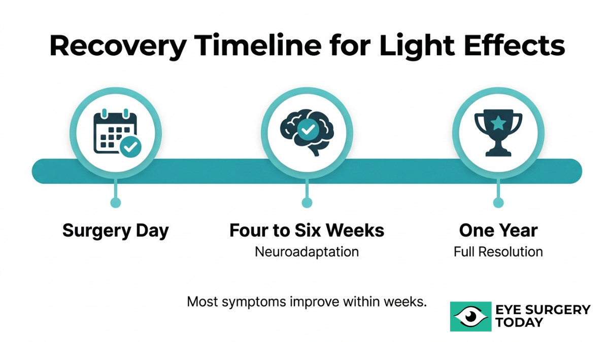
Most photic phenomena are temporary. Neuroadaptation typically reduces symptoms within four to six weeks, and persistent dysphotopsia beyond one year is uncommon. For cases that do not resolve on their own, treatments range from pharmacological pupil constriction with eye drops to surgical options such as IOL exchange, piggyback lens implantation, or reverse optic capture.
Preoperative IOL selection and a thorough discussion of expectations with your cataract surgeon remain among the most effective ways to reduce the likelihood and impact of these visual disturbances.
What Is Dysphotopsia After Cataract Surgery?
Dysphotopsia after cataract surgery is an unwanted visual disturbance caused by the interaction between light and the intraocular lens (IOL). It takes two forms: positive dysphotopsia and negative dysphotopsia.
What Is Positive Dysphotopsia?
Positive dysphotopsia is the perception of bright, unwanted light artifacts after cataract surgery. Patients typically describe these visual disturbances using terms such as “streaks,” “rays,” “arcs,” and “flickers,” according to the American Academy of Ophthalmology. These artifacts usually appear centrally or in the mid-periphery of vision, often triggered by an oblique light source hitting the edge of the IOL.
The design and material of the intraocular lens play a significant role. Square-edge IOL designs, while effective at reducing posterior capsule opacification, can reflect light onto the retina in ways that produce these bright artifacts. IOL material matters as well; silicone and copolymer lenses, such as the STAAR Surgical Collamer lens, tend to produce lower rates of positive dysphotopsia than acrylic lenses because of their reduced surface reflectivity.
For patients experiencing persistent bright artifacts, identifying the lens material and edge design is often the first step toward understanding the source of the problem.
What Is Negative Dysphotopsia?
Negative dysphotopsia is the perception of a dark shadow in the peripheral visual field after cataract surgery. According to the American Academy of Ophthalmology, patients describe it as a dark, temporal, crescent-shaped or arc-shaped shadow, sometimes compared to a “horse blinder” effect. Unlike positive dysphotopsia, this phenomenon involves an absence of light reaching a portion of the retina rather than unwanted bright artifacts.
Several risk factors can increase the likelihood of developing negative dysphotopsia:
- A small pupil size limits the light entering around the IOL optic edge.
- Large angle kappa values (the angle between the visual axis and the pupillary center) can shift the IOL’s optical zone relative to the line of sight.
- A high-index-of-refraction IOL material may create a sharper boundary between illuminated and shadowed retinal zones.
For persistent cases that do not resolve through neuroadaptation, Nd:YAG laser removal of the nasal capsule overlying the IOL can be beneficial. Negative dysphotopsia remains one of the more challenging postoperative visual complaints to manage, which makes understanding its causes and risk factors especially valuable before surgery.
With these definitions in place, recognizing what halos, glare, and starbursts actually look like helps distinguish them from dysphotopsia.
What Do Halos, Glare, and Starbursts Look Like After Cataract Surgery?
Halos, glare, and starbursts after cataract surgery look like distinct light artifacts that appear around or near light sources, especially at night. Each phenomenon has a different visual pattern.
What Do Halos Around Lights Look Like After Cataract Surgery?
Halos around lights after cataract surgery look like soft, circular rings of light that surround a point light source. They typically appear as a glowing, hazy circle extending outward from headlights, streetlamps, or other bright objects in dim conditions. The ring is usually most noticeable against a dark background, where the contrast between the bright center and the surrounding glow becomes pronounced. Patients often describe the effect as similar to looking at a light through a foggy window. Halos can vary in size and intensity depending on the type of intraocular lens implanted and the pupil’s diameter in low light. For most patients, these rings are most disruptive during nighttime driving, when oncoming headlights produce overlapping circles that reduce visual clarity.
What Does Glare Look Like After Cataract Surgery?
Glare after cataract surgery looks like a diffuse wash or veil of brightness that spreads across part of the visual field when exposed to a strong light source. Unlike halos, which form a defined ring, glare creates a broader scattering effect that can temporarily reduce contrast and make it harder to distinguish objects near the light. Patients commonly describe glare as an uncomfortable brightness or a “washed out” appearance, particularly when facing headlights, sunlight reflecting off wet pavement, or overhead lighting in indoor spaces. According to EyeWiki, published by the American Academy of Ophthalmology, positive dysphotopsia is defined as bright artifacts of light occurring centrally or mid-peripherally, typically stimulated by an oblique light source. Glare is one of the most commonly reported forms of this phenomenon. The severity often depends on lighting angle and IOL optic edge design.
What Do Starbursts Look Like After Cataract Surgery?
Starbursts after cataract surgery look like fine lines or rays of light radiating outward from a central point source, similar to a star shape. These spoke-like projections extend in multiple directions from headlights, streetlights, or other bright pinpoint sources, creating a spiky or spidering pattern. Starbursts tend to be most visible at night and are distinct from halos because the light extends outward in defined rays rather than forming a smooth ring. The number and length of the rays can vary between patients. In many cases, the diffractive or refractive properties of the implanted IOL contribute to how pronounced these patterns appear. Recognizing the specific visual pattern of starbursts can help patients describe their symptoms more accurately to their eye doctor.
Understanding how each of these photic phenomena appears can help guide conversations about IOL selection and treatment options.
What Causes Dysphotopsia After Cataract Surgery?
Dysphotopsia after cataract surgery is caused by the way light interacts with the intraocular lens (IOL), surrounding eye structures, and the pupil. The following sections cover IOL edge design, lens position, pupil size, residual capsule, and IOL material.
How Does IOL Edge Design Cause Dysphotopsia?
IOL edge design causes dysphotopsia by reflecting light at angles that create unwanted visual artifacts on the retina. Square-edge IOL designs were introduced specifically to reduce posterior capsule opacification, but this same sharp edge geometry is the primary cause of positive dysphotopsia. When light enters the eye from an oblique angle, the square edge acts as a reflective surface, directing stray light onto the retina as arcs, streaks, or crescents.
According to a clinical study published on ResearchGate, preoperative counseling about the possibility of dysphotopsia does not change its incidence but significantly improves a patient’s ability to adapt to these visual sensations postoperatively. This finding highlights why thorough preoperative discussions remain one of the most practical tools surgeons have, even when the optical cause cannot be fully eliminated through lens design alone.
Clinicians also treat dysphotopsia as a diagnosis of exclusion, meaning other potential causes such as vitreoretinal traction, posterior capsular striae, or residual astigmatism must be ruled out before attributing symptoms to IOL edge effects.
How Does IOL Position or Tilt Cause Dysphotopsia?
IOL position or tilt causes dysphotopsia by shifting the optical axis of the lens relative to the visual axis of the eye. When an IOL is decentered or tilted within the capsular bag, light rays may not focus symmetrically on the retina. This misalignment can direct stray light toward the peripheral retina, producing positive dysphotopsia symptoms such as arcs or streaks.
Tilt can also alter the gap between the IOL optic edge and the pupil margin, which may contribute to negative dysphotopsia by creating zones where light fails to reach the retina. Even subtle degrees of IOL displacement, sometimes less than a millimeter, can generate noticeable photic disturbances. Proper surgical centration and stable capsular bag fixation help minimize these positional causes.
How Does Pupil Size Contribute to Dysphotopsia?
Pupil size contributes to dysphotopsia by determining how much of the IOL optic edge is exposed to incoming light. A larger pupil allows light to reach the peripheral edges of the IOL, increasing the likelihood of edge-related reflections that cause positive dysphotopsia. Conversely, a smaller pupil may create a sharper boundary between light refracted through the IOL and light that bypasses the optic entirely, potentially worsening negative dysphotopsia.
Pupil dynamics in low-light conditions, such as nighttime driving, are particularly relevant. As the pupil dilates in dim environments, more oblique light interacts with the IOL edge, which is why many patients report halos and glare primarily at night. This relationship between pupil diameter and symptom severity is one reason pharmacological pupil constriction can be effective in managing positive dysphotopsia.
How Does Residual Lens Capsule Affect Dysphotopsia?
Residual lens capsule affects dysphotopsia by altering how light passes around and through the IOL. The anterior capsular rim, left intact after capsulorhexis, can create a secondary refractive edge that redirects light. When this rim overlaps the IOL optic too precisely, it may contribute to the illumination gap associated with negative dysphotopsia.
Posterior capsule opacification (PCO), a common postoperative change where residual lens epithelial cells proliferate on the capsule behind the IOL, can scatter light and produce symptoms resembling positive dysphotopsia. The interaction between IOL optics, angle kappa, and capsular anatomy makes the residual capsule an important variable in the overall photic disturbance profile after cataract surgery.
How Does IOL Material Affect Light Scatter?
IOL material affects light scatter through differences in refractive index and surface reflectivity. Materials with a high index of refraction, such as certain hydrophobic acrylics, reflect more light internally at the lens surfaces. This internal reflection can redirect stray light toward the retina, producing the arcs or streaks characteristic of positive dysphotopsia.
Silicone IOLs, which have a lower index of refraction, tend to produce less surface reflectivity and are associated with lower rates of positive dysphotopsia. The trade-off between material properties, optical clarity, and dysphotopsia risk is an important consideration during IOL selection, making preoperative discussion between surgeon and patient essential for setting realistic visual expectations.
Understanding these material-level causes helps clarify which IOL types may be more likely to produce specific photic phenomena.
Which Intraocular Lenses Are More Likely to Cause Dysphotopsia?
The intraocular lenses more likely to cause dysphotopsia include multifocal IOLs and extended depth of focus designs, though monofocal and toric lenses can also produce symptoms.
Do Multifocal IOLs Cause More Halos and Glare?
Multifocal IOLs can cause more halos and glare than other lens types due to their multiple focal zones, which split incoming light to provide vision at different distances. This light-splitting design inherently increases the chance of photic phenomena, particularly under low-light conditions such as nighttime driving.
According to a study published in The Open Ophthalmology Journal comparing monofocal IOLs, Clareon eyes had a higher incidence of glare, halos, and positive dysphotopsia, while AcrySof IQ eyes had a higher incidence of negative dysphotopsia. These findings highlight how even material and platform differences within the same lens category can shift the type of visual disturbance a patient experiences. For patients prioritizing spectacle independence, the trade-off between near vision convenience and potential photic side effects is one of the most important preoperative conversations to have.
Do Extended Depth of Focus IOLs Cause Dysphotopsia?
Extended depth of focus IOLs can cause dysphotopsia, though they may produce fewer distinct halos than traditional multifocal designs. EDOF lenses elongate a single focal point rather than creating separate near and far zones, which can result in a smoother visual profile. However, their diffractive optics still interact with light in ways that generate photic symptoms.
As noted by the American Academy of Ophthalmology (EyeNet), photic phenomena such as halos, glare, and starbursts are sometimes categorized as “diffractive dysphotopsias” when caused by the diffractive properties of multifocal or EDOF IOLs. While many patients tolerate EDOF lenses well, those with large pupils or high visual demands at night should discuss these potential symptoms with their surgeon before selecting this lens category.
Do Monofocal IOLs Cause Dysphotopsia?
Monofocal IOLs can cause dysphotopsia, even though they use a single focal point and lack the light-splitting optics of multifocal designs. Both positive and negative dysphotopsia have been well documented with monofocal lenses. Square-edge optic designs, while effective at reducing posterior capsule opacification, can reflect oblique light onto the retina and produce arcs, streaks, or halos. The lens material also plays a role; high-index acrylic IOLs tend to generate more internal reflections than silicone alternatives. Because monofocal IOLs remain the most commonly implanted lens worldwide, clinicians frequently encounter dysphotopsia complaints in this population, even when surgical outcomes are otherwise excellent.
Do Toric IOLs Cause Dysphotopsia?
Toric IOLs can cause dysphotopsia at rates similar to other lens platforms that share the same base optic design. A toric IOL corrects pre-existing corneal astigmatism by incorporating cylindrical power into the lens, but this astigmatism-correcting feature does not independently increase the risk of halos, glare, or starbursts. The dysphotopsia risk with a toric lens depends primarily on its underlying optic type. A toric monofocal IOL carries the same photic profile as its non-toric monofocal counterpart, while a toric multifocal lens carries the elevated risk associated with multifocal optics.
Understanding how each IOL category interacts with light can help guide the lens selection conversation with your cataract surgeon.
How Common Are Halos, Glare, and Starbursts After Cataract Surgery?
Halos, glare and light sensitivity after cataract surgery are common, affecting up to 49% of patients in the early postoperative period. The frequency varies significantly depending on the type of intraocular lens implanted and individual patient factors.
According to the American Academy of Ophthalmology (EyeNet), positive and negative dysphotopsias are reported in as many as 49% of patients in the early postoperative period after cataract surgery. Multifocal IOL recipients tend to experience higher rates: a comparative study published in PubMed Central found that 65% to 79% of patients reported halos and 43% to 64% reported starbursts six months after multifocal IOL implantation. Negative dysphotopsia, perceived as a dark crescent-shaped shadow, is estimated to occur in 15% to 26% of patients during the first week post-surgery, though it persists at one year in only 0.13% to 3.2% of cases.
These numbers, while initially striking, reflect a reality that most photic phenomena are temporary. Neuroadaptation allows the brain to gradually filter out unwanted visual artifacts for the majority of patients. The IOL type selected before surgery remains the single most influential variable in determining whether these symptoms occur and how intense they may be.
How Long Do Halos, Glare, and Starbursts Last After Cataract Surgery?
Halos, glare, and starbursts after cataract surgery typically last four to six weeks, though some patients may notice symptoms for up to a year. The subsections below cover natural resolution timelines and warning signs that symptoms may need medical attention.
Do Halos and Glare Go Away on Their Own Over Time?
Halos and glare do go away on their own over time in most cases. The brain gradually adjusts to the new intraocular lens through a process called neuroadaptation, which reduces how prominently these visual disturbances register. According to the American Academy of Ophthalmology (EyeNet), most patients experience improvement in dysphotopsia symptoms due to neuroadaptation, typically occurring within four to six weeks, though it can take up to a year.
Several factors influence how quickly symptoms resolve:
- IOL type: Multifocal and extended depth of focus lenses may produce longer-lasting photic phenomena than monofocal designs.
- Pupil size: Larger pupils allow more peripheral light to interact with the IOL edge, potentially extending symptom duration.
- Individual neural processing: Functional MRI studies have confirmed that neuroadaptation activates brain areas associated with learning and cognitive control, which helps explain why timelines vary between patients.
For most patients, patience during the early postoperative period is warranted. Persistent symptoms beyond three to six months, however, deserve a closer look with your eye doctor. Beyond these specific visual disturbances, understanding general vision after cataract surgery helps set realistic expectations for overall visual quality and adaptation.
When Should You Be Concerned That Symptoms Are Not Improving?
You should be concerned that symptoms are not improving if halos, glare, or starbursts remain unchanged or worsen beyond three to six months after cataract surgery. While neuroadaptation resolves most cases within weeks, a small percentage of patients experience persistent dysphotopsia that may require clinical evaluation.
Signs that warrant contacting your ophthalmologist include:
- Symptoms that intensify rather than gradually fade over time.
- A new dark, crescent-shaped shadow appearing in your peripheral vision, which may indicate negative dysphotopsia.
- Difficulty with night driving that does not improve, as this is strongly associated with ongoing dysphotopsia and reduced contrast sensitivity.
- Visual disturbances that interfere with daily activities such as reading or recognizing faces.
Dysphotopsia is primarily a diagnosis of exclusion, so your surgeon may need to rule out other causes such as dry eye after surgery, posterior capsule opacification, residual astigmatism, or retinal issues before determining the best course of action. Early communication with your eye care provider can help identify whether observation, pharmacological treatment, or a procedural intervention is appropriate.
Who Is More at Risk for Dysphotopsia After Cataract Surgery?
Certain patients face a higher risk of dysphotopsia after cataract surgery based on pupil characteristics, pre-existing eye conditions, and age-related factors.
Are Patients with Large Pupils More at Risk?
Patients with large pupils may be more at risk for certain types of dysphotopsia, particularly positive dysphotopsia. A larger pupil diameter allows more oblique light to reach the IOL edge, increasing the likelihood of unwanted reflections perceived as halos, arcs, or streaks. Interestingly, risk factors for negative dysphotopsia include a small pupil and large angle kappa values, meaning pupil size influences each type differently.
According to a study of north Indian patients published in PubMed Central, the incidence of negative dysphotopsia was 9%, with more than 80% of those cases being transient and resolving over time. Most patients experience symptom improvement through neuroadaptation, typically within four to six weeks, though adaptation can take up to a year. Pupil size alone does not determine outcome; the interplay between pupil diameter, IOL optic size, and individual anatomy shapes each patient’s overall risk profile.
Are Patients with Prior Eye Conditions More at Risk?
Patients with prior eye conditions are more at risk for dysphotopsia after cataract surgery. Pre-existing corneal irregularities, severe dry eye, and conditions that alter the optical surface can introduce higher-order aberrations that amplify unwanted light phenomena following IOL implantation.
Irregular corneal topography, for instance, may scatter light in unpredictable ways that compound the reflective effects of a new intraocular lens. Patients with a history of glaucoma, retinal conditions, or prior refractive surgery may also perceive photic disturbances more acutely due to changes in overall light processing. A thorough preoperative evaluation of the corneal surface and ocular history helps surgeons anticipate which patients may be more susceptible. This is one reason why a detailed eye health history matters as much as the lens selection itself when planning cataract surgery.
Does Age Affect the Risk of Dysphotopsia?
Age can affect the risk of dysphotopsia after cataract surgery, though the relationship is nuanced. Younger patients tend to have larger pupils and more responsive visual systems, which may increase sensitivity to unwanted light artifacts from IOL edges. Older patients, conversely, often have smaller pupils that naturally limit some positive dysphotopsia but may increase susceptibility to negative dysphotopsia.
Neuroadaptation also plays a role in how age influences outcomes. The brain’s ability to suppress unwanted visual signals varies with age; younger patients sometimes adapt more quickly, while older patients may require a longer adjustment period. Regardless of age, individual anatomy and IOL characteristics remain the strongest predictors. Age is best understood as one contributing variable within a broader risk assessment rather than a standalone determinant.
Understanding these risk factors can help guide the conversation with your cataract surgeon about the best approach for your specific situation.
How Is Dysphotopsia Diagnosed After Cataract Surgery?
Dysphotopsia is diagnosed after cataract surgery primarily through a process of exclusion, where the clinician rules out other treatable causes of visual disturbance before confirming the diagnosis. The evaluation combines a thorough clinical examination with the patient’s own description of symptoms.
Because no single objective test can measure dysphotopsia directly, diagnosis relies heavily on what the patient reports. Clinicians must first eliminate conditions that can mimic these symptoms, such as vitreoretinal traction, posterior capsular striae, or residual astigmatism. According to the American Academy of Ophthalmology (EyeNet), dysphotopsias are primarily a diagnosis of exclusion, requiring clinicians to rule out these alternative causes before attributing visual complaints to IOL-related photic phenomena.
The specific language a patient uses provides important diagnostic clues. Descriptions of bright artifacts, such as streaks, rays, arcs, or flickers, point toward positive dysphotopsia. Reports of a dark shadow or a “horse blinder” effect in the temporal visual field suggest negative dysphotopsia. Distinguishing between these two types guides the clinician toward the underlying mechanism and the appropriate management strategy.
Standardized patient-reported outcome measures can supplement the clinical interview. The NEI-VFQ-25 is a widely used vision-specific questionnaire that captures the subjective impact of visual disturbances on daily activities, such as reading and driving. While it does not diagnose dysphotopsia on its own, it helps quantify how significantly the symptoms affect a patient’s quality of life, which can inform treatment decisions.
A comprehensive slit-lamp examination, assessment of IOL position, and evaluation of the posterior capsule round out the diagnostic workup. In practice, the most reliable diagnostic pathway combines careful listening to the patient’s symptom description with systematic exclusion of other postoperative complications. Understanding how dysphotopsia is identified helps frame the next step: exploring available treatments.
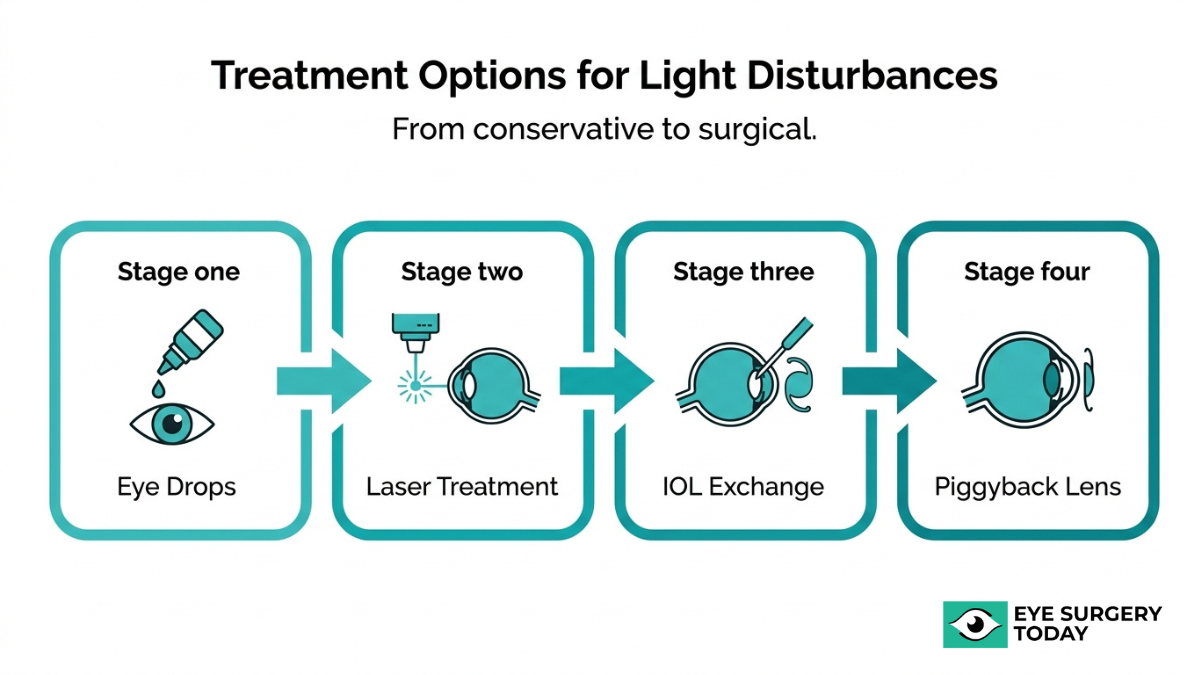
What Are the Possible Treatments for Dysphotopsia After Cataract Surgery?
The possible treatments for dysphotopsia after cataract surgery range from conservative options, such as eye drops, to surgical interventions like IOL exchange or reverse optic capture. The following subsections cover each approach.
Can Eye Drops or Medications Help Reduce Dysphotopsia?
Yes, eye drops can help reduce dysphotopsia in certain cases. Pharmacological pupil constriction using dilute pilocarpine or brimonidine drops can often reduce positive dysphotopsia symptoms by blocking light from hitting the IOL edge, according to Review of Ophthalmology. These drops work by making the pupil smaller, which limits the amount of peripheral light that interacts with the lens optic. For many patients experiencing mild to moderate glare, arcs, or streaks, this conservative approach is typically the first step an eye doctor may recommend before considering any surgical options.
Can a YAG Laser Capsulotomy Help With Dysphotopsia?
Yes, a YAG laser capsulotomy can help with dysphotopsia in specific situations. This procedure uses a focused laser to remove or alter portions of the lens capsule surrounding the intraocular lens. When posterior capsule opacification develops after cataract surgery, it may contribute to light scatter that worsens photic symptoms. In some cases of negative dysphotopsia, targeted removal of the nasal capsule overlying the IOL may reduce the shadow effect. However, not every patient with dysphotopsia is a candidate for this approach, so your ophthalmologist will evaluate whether capsular factors are contributing to your symptoms before recommending it.
Can IOL Exchange Surgery Correct Persistent Dysphotopsia?
Yes, IOL exchange surgery can correct persistent dysphotopsia when conservative treatments have failed. This procedure involves removing the original intraocular lens and replacing it with a different IOL that may have a different optic design, edge profile, or material composition. IOL exchange is generally reserved for cases where symptoms remain bothersome after several months of observation and neuroadaptation has not provided relief. Because this is a more invasive intervention than drops or laser treatment, surgeons carefully weigh the potential benefits against surgical risks before recommending it.
Can IOL Repositioning Resolve Dysphotopsia Symptoms?
Yes, IOL repositioning can resolve dysphotopsia symptoms in cases where lens misalignment contributes to unwanted light phenomena. If the intraocular lens has shifted, tilted, or decentered within the capsular bag, peripheral light rays may interact with the IOL edge differently than intended, producing glare or shadows. Repositioning the IOL back to its optimal centration may reduce these optical disturbances. This approach is less invasive than a full IOL exchange and can be effective when imaging confirms that lens position is a contributing factor.
Can a Secondary Piggyback Lens Help With Dysphotopsia?
Yes, a secondary piggyback lens can help with dysphotopsia. Piggyback IOL implantation involves placing a second intraocular lens in the ciliary sulcus, in front of the original lens. According to an AAO Editors’ Choice clinical evaluation, this technique resolved negative dysphotopsia in 8 out of 11 cases (72.7%). The additional lens may alter how light transitions between the IOL optic and the surrounding anatomy, reducing the illumination gap responsible for shadow symptoms. IOL material also plays a role; acrylic lenses with a high index of refraction are associated with higher rates of internal reflection and positive dysphotopsia compared to silicone lenses with a lower index.
Can Reverse Optic Capture Help With Negative Dysphotopsia?
Yes, reverse optic capture can help with negative dysphotopsia. This technique repositions the IOL optic anterior to the capsulorhexis edge while the haptics remain in the capsular bag. The goal is to eliminate the illumination gap on the retina, a zone between light refracted by the IOL and light that misses the optic entirely, which is hypothesized to cause the characteristic dark crescent or shadow. According to a retrospective study published by the American Academy of Ophthalmology, secondary reverse optic capture resolved negative dysphotopsia in 21 out of 22 eyes (95.5%). For patients with persistent negative dysphotopsia that has not responded to observation or conservative measures, this procedure represents one of the most effective surgical options currently available.
With treatment options ranging from simple eye drops to advanced surgical techniques, understanding how to reduce dysphotopsia risk before surgery can be equally valuable.
How Can You Reduce the Risk of Dysphotopsia Before Cataract Surgery?
You can reduce the risk of dysphotopsia before cataract surgery by selecting an appropriate IOL and having a thorough preoperative discussion with your surgeon.
How Does Choosing the Right IOL Help Prevent Dysphotopsia?
Choosing the right IOL helps prevent dysphotopsia by matching the lens design and material to your specific eye anatomy and visual needs. IOL edge design, optic diameter, and material composition all influence how light interacts with the implant after surgery.
Key IOL factors that may affect dysphotopsia risk include:
- Edge design: Round-edge optics may produce fewer internal reflections than square-edge designs.
- Material and refractive index: Silicone and copolymer lenses typically produce lower surface reflectivity than certain high-index acrylic materials.
- Optic diameter: Larger optic sizes can reduce the chance of light bypassing the lens edge.
- Lens category: Multifocal and extended depth-of-focus IOLs carry higher rates of halos and starbursts than monofocal options.
According to a comparative study published in the Journal of Cataract & Refractive Surgery, the Alcon PanOptix trifocal IOL demonstrated lower visual disturbances of starbursts and glare compared to the Johnson & Johnson Tecnis Synergy IOL. This underscores why lens selection, guided by your surgeon’s assessment of pupil size, angle kappa, and corneal health, is one of the most impactful steps you can take before surgery.
How Does Discussing Expectations With Your Surgeon Help?
Discussing expectations with your surgeon helps by preparing you to recognize and adapt to visual changes that may occur after cataract surgery. According to a clinical study published on ResearchGate, preoperative counseling regarding the possibility of dysphotopsia does not change its incidence but significantly improves the patient’s ability to adapt to the visual sensations postoperatively.
A productive presurgical conversation should cover:
- The likelihood of temporary halos, glare, or starbursts based on your chosen IOL type.
- How neuroadaptation typically reduces symptoms over weeks to months.
- Your specific visual priorities, such as night driving or reading, that may influence lens selection.
- Any pre-existing conditions like dry eye or irregular corneal topography that could heighten symptoms.
Patients who understand what to expect tend to report greater satisfaction, even when mild photic phenomena are present. Setting realistic goals before surgery remains one of the simplest yet most effective ways to improve the overall cataract surgery experience.
When Should You Contact Your Eye Doctor About Dysphotopsia?
You should contact your eye doctor about dysphotopsia when symptoms persist beyond the typical neuroadaptation window, worsen over time, or interfere with essential daily tasks. The following situations warrant a prompt call to your ophthalmologist:
- Symptoms persist beyond six weeks without improvement. Most dysphotopsia cases improve within four to six weeks as the brain adapts to the new intraocular lens. According to a report from the American Academy of Ophthalmology, neuroadaptation typically occurs within this timeframe, though it can take up to a year. If halos, glare, or starbursts remain unchanged after the initial weeks, your eye doctor can assess whether an underlying issue requires attention.
- A new dark shadow or crescent appears in your peripheral vision. Negative dysphotopsia produces a temporal, arc-shaped shadow that patients sometimes describe as a “horse blinder” effect. While often transient, a persistent or expanding dark area should be evaluated to rule out other conditions, such as vitreoretinal traction or retinal complications.
- Visual disturbances suddenly worsen after a period of stability. Gradual improvement followed by a sudden increase in light artifacts may indicate posterior capsule opacification or a shift in IOL position. Both conditions are treatable, but early detection matters.
- Night driving becomes unsafe. Difficulty driving at night is strongly associated with dysphotopsia and reduced contrast sensitivity. If halos or starbursts around oncoming headlights compromise your ability to drive safely, your ophthalmologist may recommend interventions such as dilute pilocarpine drops or further evaluation.
- Symptoms cause significant anxiety or reduce your quality of life. Even when visual acuity tests appear normal, dysphotopsia can meaningfully affect daily comfort. Your eye doctor can discuss management options ranging from pharmacological approaches to surgical interventions for persistent cases.
A reasonable guideline: mild, stable symptoms that gradually lessen over the first month rarely need urgent intervention. However, any worsening pattern, new symptoms, or functional limitation deserves clinical evaluation. Early communication with your surgeon allows timely diagnosis and, when necessary, access to effective treatments before symptoms become entrenched.
Understanding when to seek help is important, but so is recognizing how these visual disturbances may affect your everyday routine.
How Can Dysphotopsia Affect Daily Activities Like Driving at Night?
Dysphotopsia can affect daily activities like driving at night by introducing unwanted visual disturbances, such as halos, glare, and starbursts around oncoming headlights, streetlights, and traffic signals. These photic phenomena may reduce contrast sensitivity and make it harder to judge distances or read road signs in low-light conditions.
According to EyeWiki (American Academy of Ophthalmology), the NEI-VFQ-25 is a widely used vision-specific patient-reported outcome measure that captures the subjective impact of vision on daily activities like reading and driving. This type of validated tool helps clinicians understand how significantly dysphotopsia symptoms interfere with a patient’s routine.
Beyond driving, tasks that involve transitioning between bright and dim environments, such as walking into a darkened theater or navigating a dimly lit parking garage, can also become more challenging. Reading under certain lighting conditions and using screens at night may feel uncomfortable when halos or streaks persist in the visual field.
For many patients, the functional impact of dysphotopsia is more disruptive than any measurable change in visual acuity alone. A patient may test at 20/20 on a standard eye chart yet still struggle with nighttime driving because standard acuity measurements do not capture the quality of vision under real-world conditions. Discussing these specific activity-related concerns with your eye doctor can help guide both IOL selection before surgery and management strategies afterward.
How Can Eye Surgery Today Help You Understand Dysphotopsia After Cataract Surgery?
Eye Surgery Today can help you understand dysphotopsia after cataract surgery by providing surgeon-reviewed educational guides that explain visual symptoms, IOL selection, and what to expect during recovery.
Can Eye Surgery Today’s Surgeon-Reviewed Guides Help You Choose the Right IOL?
Yes, Eye Surgery Today’s surgeon-reviewed guides can help you choose the right IOL by breaking down how different lens designs affect postoperative visual quality. IOL selection is one of the most important decisions in cataract surgery, and understanding how lens type relates to dysphotopsia risk can make that choice more informed. Eye Surgery Today covers the differences between monofocal, multifocal, trifocal, and extended depth of focus IOLs, including how each design may influence the likelihood of halos, glare, and starbursts. Pre-existing corneal conditions, such as irregular topography or severe dry eye, can also induce aberrations that patients perceive as positive dysphotopsia after surgery, which makes preoperative evaluation and IOL matching even more critical. Eye Surgery Today presents this information in accessible, jargon-free language so patients can discuss options confidently with their surgeon.
What Are the Key Takeaways About Dysphotopsia, Halos, Glare, and Starbursts After Cataract Surgery?
The key takeaways about dysphotopsia, halos, glare, and starbursts after cataract surgery are:
- Dysphotopsia refers to unwanted visual images after cataract surgery, divided into positive types (bright artifacts such as arcs, streaks, starbursts, rings, or halos) and negative types (dark shadows or crescents).
- These symptoms are common. According to the American Academy of Ophthalmology, dysphotopsias are reported in as many as 49% of patients in the early postoperative period.
- Most cases improve with neuroadaptation over weeks to months, and persistent symptoms beyond one year are rare.
- IOL design plays a significant role. In a comparative study published in PubMed Central, 65% to 79% of patients reported halos and 43% to 64% reported starbursts six months after multifocal IOL implantation.
- Difficulty with night driving is strongly associated with dysphotopsia and reduced contrast sensitivity.
- Multiple treatment options exist for persistent cases, ranging from eye drops to surgical interventions.
Knowing what to expect before and after surgery remains the best way to manage these visual symptoms. Eye Surgery Today provides the surgeon-reviewed resources patients need to navigate every stage of this process with confidence.


