Cataract surgery is a procedure that removes the eye’s clouded natural lens and replaces it with a clear artificial intraocular lens (IOL) to restore functional vision. Results vary by individual, though the procedure may significantly improve visual clarity, color perception, and overall quality of life for most patients.
This guide covers pre-surgical vision changes, how outcomes are measured, what to expect during and after the procedure, how lens selection shapes results, factors that influence recovery, and potential complications.
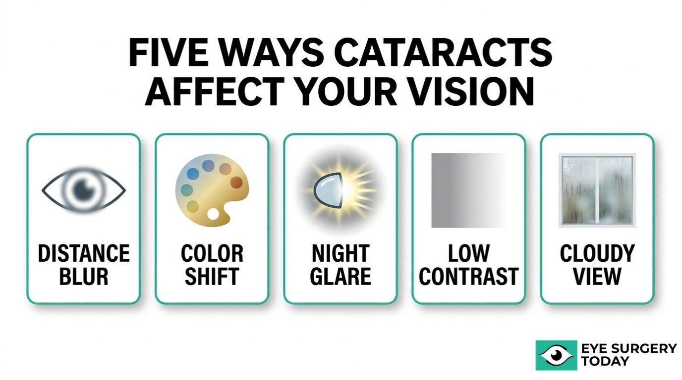
Before surgery, cataracts progressively degrade vision across multiple dimensions. Distance acuity declines, colors shift toward yellow or brown tones, night driving becomes difficult due to halos and glare, and contrast sensitivity drops across all spatial frequencies. Many patients describe their sight as looking through a foggy window.
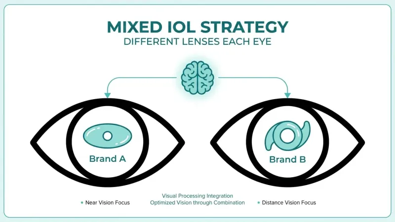
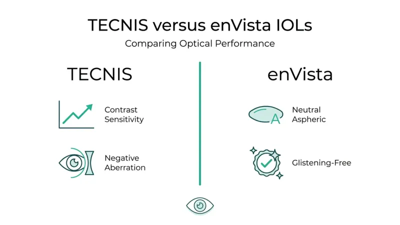
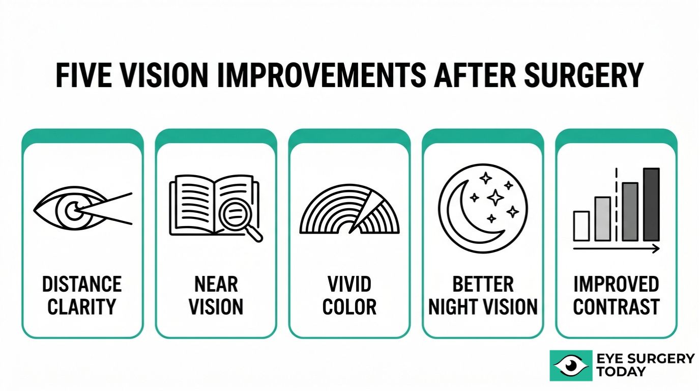
Post-surgical vision improvements may include sharper distance clarity, more vivid color perception, reduced glare sensitivity, and restored contrast. The type of IOL implanted plays a central role in determining which distances benefit most, whether near, intermediate, or far. Monofocal, multifocal, toric, and extended depth of focus lenses each offer distinct visual trade-offs.
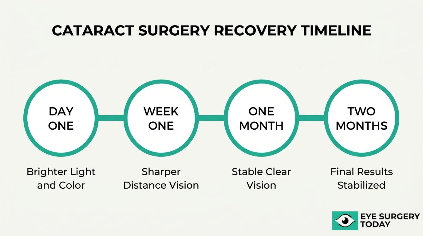
Recovery follows a general timeline: noticeable improvement often begins within hours, with progressive sharpening over the first week. Full stabilization of refraction and visual acuity typically takes four to eight weeks, during which consistent post-operative care supports the best possible outcome.
Individual factors, including pre-existing eye conditions, cataract severity, and age, can influence final results. Complications such as posterior capsule opacification or dry eye may temporarily affect vision quality but are generally treatable when detected early.
What Is Vision Like Before Cataract Surgery?
Vision before cataract surgery is typically degraded across multiple dimensions, including sharpness, color accuracy, night visibility, contrast, and overall clarity. The following subsections cover how cataracts affect distance vision, color perception, night vision, contrast sensitivity, and the characteristic blurry or cloudy appearance patients often describe.
How Do Cataracts Affect Distance Vision?
Cataracts affect distance vision by progressively scattering and blocking light as it passes through the clouded natural lens, making distant objects appear increasingly blurred. Road signs, faces across a room, and television screens may all lose definition as the opacity worsens. According to a study published in the National Institutes of Health (PMC), increasing cataract severity leads to a significant decrease in visual acuity and a loss of contrast sensitivity across all spatial frequencies. This decline often happens gradually, so many patients unconsciously compensate by squinting, moving closer to objects, or increasing screen brightness. For most people, the slow progression of distance vision loss is what ultimately prompts a conversation about surgical options.
How Do Cataracts Change Color Perception?
Cataracts change color perception by filtering light unevenly through the yellowing or browning lens, causing colors to appear faded, washed out, or shifted toward warm amber tones. Whites may take on a yellowish cast, and blues can become harder to distinguish from greens or purples. Nuclear sclerotic cataracts, the most common subtype, show the most profound impact on color perception. Many patients do not realize how much color fidelity they have lost until after surgery restores a clearer optical path. This gradual shift is particularly noticeable in daily tasks that depend on accurate color discrimination, such as matching clothing, reading color-coded labels, or appreciating artwork.
How Do Cataracts Impact Night Vision and Glare?
Cataracts impact night vision and glare by scattering incoming light across irregular opacities in the lens, creating halos around headlights, starbursts from streetlamps, and an overall reduction in low-light visibility. Driving at night often becomes one of the first activities patients report avoiding. According to a 2022 study published in Ophthalmology Science, functional visual impairment from cataracts includes deficits in visual acuity, spatial contrast sensitivity, and increased glare disability under both photopic and mesopic conditions. Oncoming headlights can feel blinding, while dimly lit environments become difficult to navigate safely. This combination of glare sensitivity and reduced night acuity represents one of the most functionally limiting aspects of pre-surgical cataract vision.
How Does Contrast Sensitivity Decline Before Surgery?
Contrast sensitivity declines before surgery because the cataract scatters light within the eye, reducing the optical system’s ability to distinguish subtle differences between an object and its background. Low-contrast situations become particularly challenging:
- Reading gray text on a white page grows difficult.
- Navigating stairs with uniform coloring becomes a fall risk.
- Facial expressions across a room lose definition.
Even when a standard letter chart still shows acceptable acuity, real-world visual performance often suffers. Contrast sensitivity testing captures this functional decline that a basic Snellen acuity measurement may miss, which is why many surgeons consider it an important part of the pre-operative evaluation.
What Does Blurry or Cloudy Vision From Cataracts Look Like?
Blurry or cloudy vision from cataracts looks like viewing the world through a foggy or frosted window. The entire visual field takes on a hazy quality rather than producing a single blurred spot. Common descriptions from patients include:
- A persistent film or fog over everything, regardless of lighting.
- Dimmed overall brightness, as if indoor lights are never quite bright enough.
- Frequent need to clean eyeglasses that are already clean.
- Double images or ghost images in the affected eye.
The severity varies with the type and location of the cataract. Posterior subcapsular cataracts, for example, can cause significant glare and reading difficulty even at relatively early stages, while nuclear cataracts tend to produce a more gradual, generalized haze. Understanding what pre-surgical vision looks and feels like helps set realistic expectations for the improvements cataract surgery may provide.
How Is Vision Typically Measured Before Cataract Surgery?
Vision before cataract surgery is typically measured using a combination of standardized tests that assess visual acuity, contrast sensitivity, glare disability, and color perception. These measurements establish a baseline and help determine surgical timing.
Ophthalmologists rely on the Snellen eye chart as the primary tool for measuring distance visual acuity, recorded as a fraction such as 20/40 or 20/80. However, visual acuity alone does not capture the full scope of cataract-related vision loss. Functional visual impairment from cataracts includes deficits in spatial contrast sensitivity and increased glare disability under both bright and dim lighting conditions, according to research published in Ophthalmology Science. Contrast sensitivity testing reveals how well the eye distinguishes objects from their background at varying spatial frequencies, while glare testing simulates real-world conditions like oncoming headlights. Color vision assessments can identify shifts in perception, particularly with nuclear cataracts. The Lens Opacities Classification System III (LOCS III) grades the cataract itself across four categories: nuclear opalescence, nuclear color, cortical, and posterior subcapsular. Together, these measurements give the surgical team a comprehensive picture of how much the cataract affects daily function, not just letter recognition on a chart.
Understanding how your baseline vision is documented helps set realistic expectations for the improvements cataract surgery may provide.
Who Is a Good Candidate for Cataract Surgery?
A good candidate for cataract surgery is someone whose lens opacity causes meaningful visual impairment that interferes with daily activities. Candidacy depends on symptom severity, overall eye health, and individual visual goals.
Most ophthalmologists consider surgery when cataracts reduce quality of life rather than relying on a single visual acuity threshold. A patient who struggles with driving at night, reading, or recognizing faces due to lens clouding may benefit from surgical intervention, even if standard distance acuity still tests at moderate levels. Functional deficits in contrast sensitivity and glare disability, not just Snellen chart readings, often guide the decision.
Several factors typically support candidacy:
- Visually significant symptoms: Blurred or cloudy vision, increased glare sensitivity, faded color perception, or difficulty with low-light tasks that corrective lenses no longer adequately address.
- Impact on daily function: Trouble driving, reading, working, or performing hobbies safely due to progressive lens opacity.
- Stable general eye health: Absence of uncontrolled conditions, such as advanced glaucoma or active diabetic retinopathy, that could limit visual improvement after surgery.
- Realistic expectations: Understanding that outcomes depend on factors like pre-existing eye conditions, IOL selection, and individual healing response.
- Medical fitness for surgery: No systemic conditions that would make sedation or the procedure itself unsafe.
Patients with coexisting eye diseases are not automatically excluded; however, their anticipated visual gains may be more limited. An ophthalmologist can help determine whether the cataract is the primary cause of vision loss or if another condition plays a larger role. According to a large-scale analysis of 368,256 cataract surgeries reported by MDinteractive, corrected distance visual acuity of 20/40 or better was achieved in 94.3% of cases, which illustrates the generally favorable outcomes when candidates are appropriately selected.
Timing also matters. Waiting until a cataract becomes extremely dense can increase surgical complexity and slightly raise complication risk. For most patients, pursuing surgery once symptoms meaningfully affect daily life strikes the best balance between benefit and procedural safety. Understanding the surgical procedure itself can help candidates feel more prepared for what comes next.
What Should You Expect During the Cataract Surgery Procedure?
You should expect a brief outpatient procedure that removes your clouded natural lens and replaces it with an artificial intraocular lens (IOL). The surgery typically involves numbing eye drops, a small corneal incision, ultrasonic lens removal, and IOL implantation.
Before the procedure begins, your surgical team will administer dilating drops to widen the pupil and anesthetic drops to numb the eye. Most patients remain awake throughout, though mild sedation may be offered to reduce anxiety. The surgeon creates a small incision in the cornea, usually between 2 and 3 millimeters, then uses ultrasonic energy through a technique called phacoemulsification to break apart and gently suction away the clouded lens. Once the natural lens is removed, a carefully selected IOL is folded, inserted through the same micro-incision, and positioned within the lens capsule.
The entire process generally takes 15 to 20 minutes per eye. Because the incision is so small, stitches are rarely needed; the wound typically self-seals. Your IOL choice significantly influences your visual outcome after surgery. According to a meta-analysis published in a 2023 review indexed in PubMed, at least 90% of patients receiving trifocal IOLs can expect to achieve complete spectacle independence. For patients prioritizing reduced dependence on glasses at multiple distances, discussing premium IOL options with a surgeon before the procedure is one of the most impactful decisions in the entire process.
Understanding how the procedure works sets the stage for knowing what vision improvements may follow in the days and weeks ahead.
What Are the Potential Vision Results After Cataract Surgery?
The potential vision results after cataract surgery may include improved distance clarity, enhanced near vision, brighter color perception, reduced glare sensitivity, and better contrast sensitivity. The following subsections cover each of these outcomes.
How May Distance Vision Improve After Cataract Surgery?
Distance vision may improve significantly after cataract surgery, with most patients achieving functional clarity for driving, reading signs, and recognizing faces at a distance. In a large-scale study of 368,256 cataract surgeries, corrected distance visual acuity (CDVA) of 20/40 or better was achieved in 94.3% of cases, according to MDinteractive’s MIPS Quality Measure data.
Advanced intraocular lens options can push these results further. Approximately 62% of eyes implanted with extended-range-of-vision IOLs achieve uncorrected distance visual acuity of 20/20 or better at 12 months post-surgery. The degree of improvement depends on factors such as IOL selection, pre-existing eye conditions, and overall ocular health. For most patients, whose mean age at the time of surgery is approximately 71.5 years, the restoration of sharp distance vision represents one of the most noticeable quality-of-life gains following the procedure.
How May Near Vision Change After Cataract Surgery?
Near vision may change after cataract surgery depending on the type of intraocular lens implanted. A standard monofocal IOL typically optimizes distance vision, which means reading glasses may still be needed for close-up tasks such as reading, sewing, or using a smartphone. Multifocal and trifocal IOLs, by contrast, are designed to provide functional near vision alongside distance correction.
It is worth noting that ocular surface changes can affect perceived near vision quality in the weeks following surgery. According to a 2024 study published in PubMed, nearly 20% of patients develop dry eye syndrome after cataract surgery, with 8.3% requiring nonroutine follow-up care. Dry eye can cause intermittent blurring that mimics poor near focus. Discussing both IOL options and post-operative dry eye management with a healthcare provider can help set realistic expectations for near vision outcomes.
How May Color and Brightness Perception Improve After Surgery?
Color and brightness perception may improve noticeably after cataract surgery because the clouded natural lens is replaced with a clear artificial intraocular lens. Cataracts, particularly nuclear sclerotic types, gradually filter out blue and violet wavelengths, giving the world a yellowish or brownish tint that worsens over time. Many patients do not realize how much color has faded until after their procedure.
Once the opacified lens is removed, the full visible light spectrum can reach the retina without obstruction. Colors often appear more vivid and saturated, and whites look brighter rather than dingy. Indoor lighting and outdoor scenery may seem remarkably more vibrant within the first few days of recovery. This restoration of accurate color perception is one of the most immediately appreciated changes patients report, even though it receives less clinical attention than visual acuity measurements.
How May Night Vision and Glare Sensitivity Change After Surgery?
Night vision and glare sensitivity may change after cataract surgery, typically improving as the opacified lens is replaced with a clear IOL. Cataracts scatter incoming light, which creates halos around headlights, starbursts from streetlamps, and overall difficulty seeing in low-light conditions. Removing the cataract eliminates this primary source of light scatter.
However, some patients experience temporary glare, halos, or light sensitivity during the early recovery period as the eye adjusts to the new lens. These symptoms, often called dysphotopsias, tend to diminish within weeks to months for most individuals. The IOL type also matters: multifocal lenses may produce more noticeable halos at night than monofocal designs. Patients who frequently drive at night should discuss lens selection carefully with their surgeon to balance daytime visual range against nighttime optical quality.
How May Contrast Sensitivity Improve After Cataract Surgery?
Contrast sensitivity may improve after cataract surgery because the procedure removes the lens opacity that degrades the eye’s ability to distinguish objects from their background. According to a study published by the National Institutes of Health (PMC), increasing cataract severity leads to a significant decrease in visual acuity and a loss of contrast sensitivity across all spatial frequencies.
Before surgery, patients often struggle to perceive subtle differences between similar shades, making tasks such as navigating stairs, reading low-contrast text, and driving in fog or rain more difficult. Once the clouded lens is replaced, light passes through the clear IOL without scattering, which restores the eye’s capacity to detect fine tonal differences. Improved contrast sensitivity often proves just as meaningful for daily functioning as sharper visual acuity, particularly for older adults navigating environments with uneven lighting.
With a clearer picture of potential post-surgical outcomes, understanding the recovery timeline helps set realistic expectations for when these improvements take shape.
How Soon After Cataract Surgery Can You Notice Vision Changes?
Vision changes after cataract surgery can begin within hours, though the timeline varies. The subsections below cover what to expect during the first day, first week, one month, and when final results typically stabilize.
What Vision Changes Are Common in the First Day After Surgery?
Vision changes common in the first day after surgery include brighter light perception, improved color vibrancy, and a general sense of clearer sight. Many patients notice these shifts within just a few hours of the procedure, even while mild blurriness or haziness persists.
Some temporary visual disturbances are also normal during this initial period:
- Mild blurriness as the eye adjusts to the new intraocular lens
- Light sensitivity or a slight glare effect in bright environments
- A faint halo appearance around lights
- Watery or slightly irritated vision from post-operative inflammation
These early effects result from corneal swelling and residual inflammation, both of which typically begin subsiding within hours. The operated eye may feel noticeably different from the untreated eye if surgery was performed on one side only. Most patients can distinguish meaningful improvement in brightness and clarity by the end of the first day, even before swelling fully resolves.
What Vision Changes Are Common in the First Week After Surgery?
Vision changes common in the first week after surgery include progressively sharper distance vision, reduced haziness, and continued improvement in color and brightness perception. According to clinical guidelines from Greenwich Eye, significant visual improvement is often noticed within the first 24 to 48 hours, though full stabilization of refraction and visual acuity typically takes 4 to 8 weeks.
During this period, fluctuating vision is normal. Clarity may shift throughout the day as the cornea heals and inflammation gradually decreases. Mild dryness and intermittent light sensitivity may still be present, though both tend to lessen as prescribed anti-inflammatory and antibiotic eye drops take effect. By the end of the first week, most patients experience a noticeable reduction in the cloudy or washed-out quality that characterized their pre-surgical vision. Consistent use of post-operative drops and avoiding eye strain can support steady progress during this phase.
What Vision Changes Are Common One Month After Surgery?
Vision changes common one month after surgery include substantially clearer distance vision, more stable refraction, and near-complete resolution of post-operative blurriness. By this point, corneal swelling has typically resolved, and the intraocular lens has settled into its permanent position.
Color perception often appears more vivid and accurate compared to pre-surgical vision, since the clouded natural lens is no longer filtering light. Contrast sensitivity also tends to improve, making it easier to distinguish objects in varied lighting conditions. Some patients may still notice mild dryness or occasional visual fluctuations, particularly in the morning or after extended screen use. For those who require updated glasses or contact lens prescriptions, eye care providers often wait until at least the one-month mark before finalizing measurements, since minor refractive shifts can still occur during this window.
When Do Final Vision Results Typically Stabilize?
Final vision results typically stabilize between 4 and 8 weeks after cataract surgery. During this window, refraction measurements become consistent enough for an accurate glasses or contact lens prescription if one is needed.
Several factors can influence the exact timeline:
- The type of intraocular lens implanted
- Pre-existing conditions such as dry eye or corneal irregularities
- The degree of post-operative inflammation experienced
- Individual healing rates, which vary by patient
In some cases, minor adjustments in visual clarity may continue for up to three months, particularly for patients with multifocal or extended depth of focus lenses that require a neuroadaptation period. Attending all scheduled follow-up appointments allows the surgeon to monitor healing and address any concerns before they affect long-term outcomes. For most patients, the vision they experience at the two-month mark closely reflects their permanent result.
With the recovery timeline understood, choosing the right intraocular lens type plays a significant role in shaping those final outcomes.
How Do Vision Results Differ by Intraocular Lens Type?
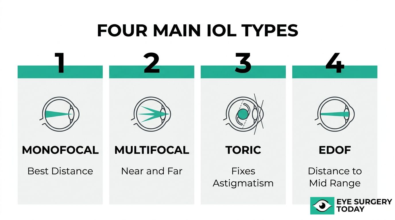
Vision results differ by intraocular lens type based on each lens design’s focal range and correction capabilities. The sections below cover monofocal, multifocal, toric, and extended depth of focus IOLs.
How May Vision Results Differ With a Monofocal IOL?
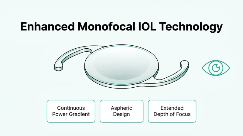
Vision results with a monofocal IOL may differ because this lens provides sharp focus at one preset distance, typically far. Most patients who receive a standard monofocal IOL achieve excellent corrected distance vision but still need reading glasses for near and intermediate tasks. According to a study published in Nature (Eye), enhanced monofocal IOLs provide significantly better intermediate-distance spectacle independence compared to standard monofocals, with an odds ratio of 7.85 (95% CI: 4.08–15.09). This makes newer enhanced monofocal designs a practical middle ground for patients who want reduced glasses dependence without the visual compromises sometimes associated with multifocal optics.
How May Vision Results Differ With a Multifocal IOL?
Vision results with a multifocal IOL may differ because this lens splits incoming light into multiple focal points, enabling clear vision at near, intermediate, and far distances simultaneously. For many patients, a multifocal IOL can significantly reduce or eliminate the need for glasses after cataract surgery. However, because light is divided across focal zones, some individuals may notice halos or glare around lights at night. These visual phenomena tend to diminish as the brain adapts over several weeks. Patients with high visual demands at multiple distances often find multifocal IOLs especially beneficial, though candidacy depends on overall eye health and lifestyle needs.
How May Vision Results Differ With a Toric IOL?
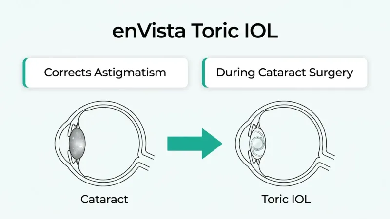
Vision results with a toric IOL may differ because this lens is specifically designed to correct pre-existing corneal astigmatism during cataract surgery. Astigmatism causes light to focus unevenly on the retina, producing blurred or distorted vision at all distances. A toric IOL counteracts this irregularity through built-in cylindrical correction aligned to the axis of astigmatism. Precise surgical placement is critical; even slight rotation of the lens can reduce its corrective effectiveness. For patients with moderate to significant astigmatism, a toric IOL can deliver noticeably sharper uncorrected distance vision compared to a standard monofocal lens. Reading glasses may still be needed for near tasks unless the toric design is combined with multifocal optics.
How May Vision Results Differ With an Extended Depth of Focus IOL?
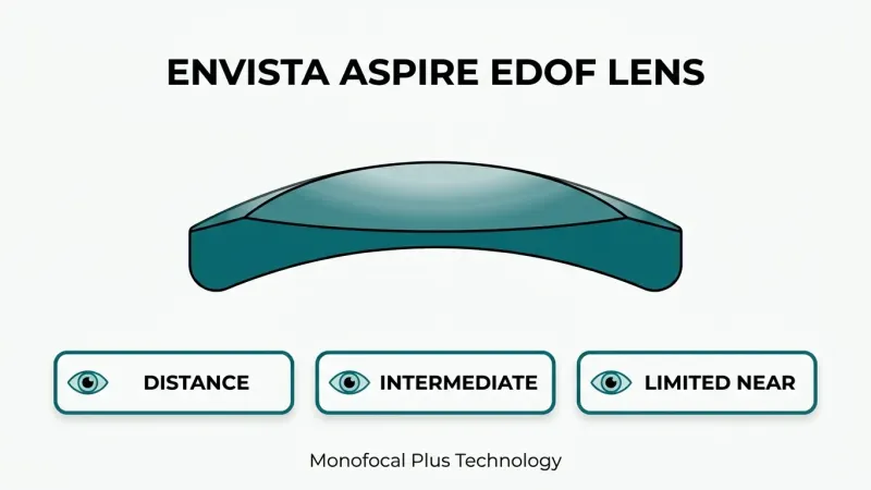
Vision results with an extended depth of focus IOL may differ because this lens elongates a single focal point rather than splitting light into separate zones. According to research published in Nature (Eye), EDOF IOLs provide superior binocular uncorrected intermediate visual acuity compared to standard monofocal lenses. This continuous range of focus covers distance and intermediate vision smoothly, which benefits activities such as computer work and dashboard reading. Because EDOF lenses avoid the multiple focal splits of multifocal designs, they typically produce fewer halos and less glare at night. Near vision may still require reading glasses for fine print, but the overall visual experience tends to feel more natural across daily tasks.
With each IOL type offering distinct visual trade-offs, other individual factors also play a role in final outcomes.
What Factors Can Affect Your After Surgery Vision Results?
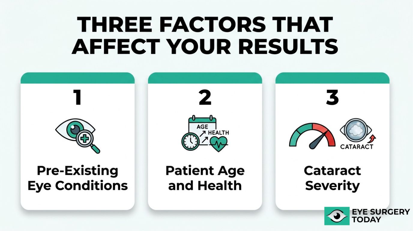
Several factors can affect your after surgery vision results, including pre-existing eye conditions, age, and the severity of your cataract at the time of the procedure.
How May Pre-Existing Eye Conditions Affect Results?
Pre-existing eye conditions may affect results by limiting the visual improvement cataract surgery can achieve. Conditions such as glaucoma, macular degeneration, and diabetic retinopathy can reduce the eye’s ability to process a clear image, even after a clouded lens has been replaced with a new intraocular lens.
Corneal irregularities, including keratoconus or corneal scarring, may also influence refractive outcomes. Because these conditions affect structures beyond the lens itself, removing the cataract addresses only one component of the visual pathway. Patients with unmanaged ocular surface disease may experience fluctuating vision that masks the true benefit of surgery.
A thorough pre-operative evaluation helps identify these conditions so that expectations can be set accurately. In many cases, treating or stabilizing a co-existing condition before surgery can improve the likelihood of a favorable outcome.
How May Age Influence Cataract Surgery Outcomes?
Age may influence cataract surgery outcomes through its effect on overall ocular health and healing capacity. According to a 2024 study published in Nature Scientific Reports, the mean age for patients undergoing cataract surgery is approximately 71.5 years, with a standard deviation of 10.7 years.
Older patients are more likely to have concurrent age-related conditions, such as macular degeneration or reduced tear production, that can limit post-operative visual gains. Healing times may also extend slightly in older individuals, though the procedure itself remains safe across a wide age range.
Younger patients who develop cataracts, often from trauma or prolonged steroid use, typically have fewer co-existing ocular conditions. This generally allows for a more predictable visual recovery. Regardless of age, candidacy is determined by the individual’s overall eye health rather than a specific age threshold.
How Does Cataract Severity at the Time of Surgery Affect Results?
Cataract severity at the time of surgery can affect results by influencing both the complexity of the procedure and the recovery timeline. A more advanced cataract typically requires greater ultrasonic energy during phacoemulsification to break up the hardened lens, which may increase corneal stress and extend healing.
Patients with dense, mature cataracts often experience more dramatic improvements in visual clarity after surgery, simply because their pre-operative vision was significantly impaired. However, prolonged cataract progression can sometimes be associated with complications such as zonular weakness, making the procedure technically more demanding.
Early-to-moderate cataracts generally involve a smoother surgical process and a faster stabilization period. According to Greenwich Eye’s clinical guidelines, full stabilization of refraction and visual acuity typically takes four to eight weeks. Addressing cataracts before they become highly advanced may support a more predictable recovery and reduce procedural risk.
With these factors in mind, understanding whether you may still need corrective lenses completes the picture of post-surgical expectations.
Will You Still Need Glasses After Cataract Surgery?
Whether you still need glasses after cataract surgery depends primarily on the type of intraocular lens (IOL) implanted during the procedure. Monofocal, multifocal, toric, and extended depth of focus lenses each produce different levels of spectacle independence for distance, intermediate, and near vision.
Most patients experience a significant reduction in glasses dependence, though complete freedom from corrective eyewear is not guaranteed for everyone. The outcome hinges on several factors: IOL selection, the accuracy of preoperative measurements, pre-existing eye conditions, and individual healing patterns.
Monofocal IOLs, the most commonly implanted lens type, typically correct distance vision effectively. However, patients with standard monofocal lenses usually still need reading glasses for close-up tasks. Enhanced monofocal designs have improved this limitation considerably. According to a 2024 meta-analysis published in Nature Eye, enhanced monofocal IOLs provide significantly better intermediate-distance spectacle independence compared to standard monofocals, with an odds ratio of 7.85.
For patients seeking broader spectacle freedom, premium lens options may reduce or eliminate the need for glasses across multiple distances:
- Multifocal IOLs split light into distinct focal points for near, intermediate, and distance vision, though some patients notice halos or glare in low-light conditions.
- Toric IOLs correct pre-existing astigmatism, which standard lenses cannot address, improving unaided distance clarity for patients with significant corneal irregularity.
- Extended depth of focus (EDOF) IOLs provide a continuous range of vision from distance through intermediate, with fewer visual disturbances than traditional multifocal designs.
Realistic expectations matter more than lens marketing. Even with advanced IOL technology, some patients may still prefer light reading glasses for prolonged close work or fine print. Your ophthalmologist can help determine which lens type best matches your visual priorities, lifestyle demands, and ocular health profile. Discussing these goals before surgery is one of the most important steps toward achieving the spectacle independence you are hoping for.
Understanding lens-related outcomes helps clarify expectations, but potential complications can also influence final vision quality.
What Are the Possible Risks or Complications That May Affect Vision?
The possible risks or complications that may affect vision after cataract surgery include posterior capsule opacification, cystoid macular edema, dry eye syndrome, and postoperative infection. Most complications are treatable when detected early.
What Is Posterior Capsule Opacification After Cataract Surgery?
Posterior capsule opacification (PCO) is a condition where the thin membrane behind the intraocular lens becomes cloudy after cataract surgery. Often called a “secondary cataract,” PCO occurs when residual lens epithelial cells migrate and proliferate across the posterior capsule, gradually blurring vision.
According to a study published in Nature (Eye), the overall incidence of PCO ranges from 7.1% to 22.6% at five years post-surgery, depending on the IOL material and design. Symptoms may include progressively hazy vision, increased glare sensitivity, and reduced contrast, often resembling the original cataract symptoms.
A quick, painless outpatient procedure called YAG laser capsulotomy can treat PCO effectively. The laser creates a small opening in the clouded capsule, restoring clear vision within days. Because PCO develops gradually over months or years, patients who notice a slow decline in visual clarity after an initially successful surgery should alert their eye care provider promptly.
What Are the Signs of Cystoid Macular Edema After Surgery?
The signs of cystoid macular edema after surgery include blurred or distorted central vision, difficulty reading, and a washed-out appearance to colors. Pseudophakic cystoid macular edema (PCME) involves fluid accumulation in the macula, the retina’s central zone responsible for sharp detail.
According to a study published by the National Institutes of Health, the incidence of postoperative PCME is estimated at 5% after uncomplicated cataract surgery. Symptoms typically emerge four to six weeks after the procedure, which can catch patients off guard since initial recovery may have seemed normal.
Treatment usually involves a combination of topical anti-inflammatory eye drops, including nonsteroidal anti-inflammatory drugs (NSAIDs) and corticosteroids. Most cases resolve with consistent medical therapy, though persistent PCME may require additional interventions. Early detection through routine postoperative visits remains the most reliable way to manage this complication before it causes lasting visual changes.
How May Dry Eye Affect Vision Quality After Cataract Surgery?
Dry eye may affect vision quality after cataract surgery by causing fluctuating blurriness, a gritty or burning sensation, and increased light sensitivity. The surgical incision and microscope light exposure can temporarily disrupt the corneal nerves that regulate tear production, leading to an unstable tear film.
Nearly 20% of patients develop dry eye syndrome following cataract surgery, with 8.3% requiring nonroutine follow-up care, according to research published in the National Institutes of Health (PubMed). An unstable tear film scatters light unevenly across the cornea, which may reduce the sharpness of vision even when the IOL itself is functioning perfectly.
Management options include:
- Preservative-free artificial tears used frequently throughout the day.
- Prescription anti-inflammatory drops to reduce ocular surface inflammation.
- Punctal plugs to help retain moisture on the eye’s surface.
- Warm compresses and lid hygiene to support healthy meibomian gland function.
Pre-existing dry eye deserves attention before surgery, since addressing it early can improve both comfort and visual outcomes.
What Is the Risk of Infection or Inflammation After Surgery?
The risk of infection after cataract surgery is low but clinically significant. Endophthalmitis, a severe intraocular infection, occurs at an overall rate of approximately 0.09% within 42 days of intraocular surgery, according to the American Academy of Ophthalmology.
Warning signs that may indicate infection or significant inflammation include:
- Sudden, severe eye pain that worsens rather than improves.
- Rapid vision loss or increasing haziness days after surgery.
- Pronounced redness or swelling around the eye.
- Increased sensitivity to light beyond normal postoperative levels.
Surgeons typically prescribe prophylactic antibiotic and anti-inflammatory eye drops before and after the procedure to minimize this risk. Strict adherence to the prescribed drop schedule, avoiding touching the eye, and keeping water away from the surgical site during early recovery all help reduce the chance of infection. Any sudden change in comfort or clarity within the first few weeks warrants immediate medical evaluation, since early treatment of endophthalmitis can be critical to preserving vision.
Understanding these potential complications helps set realistic expectations for the broader recovery process.
What Can You Do to Support the Best Possible Vision Recovery?
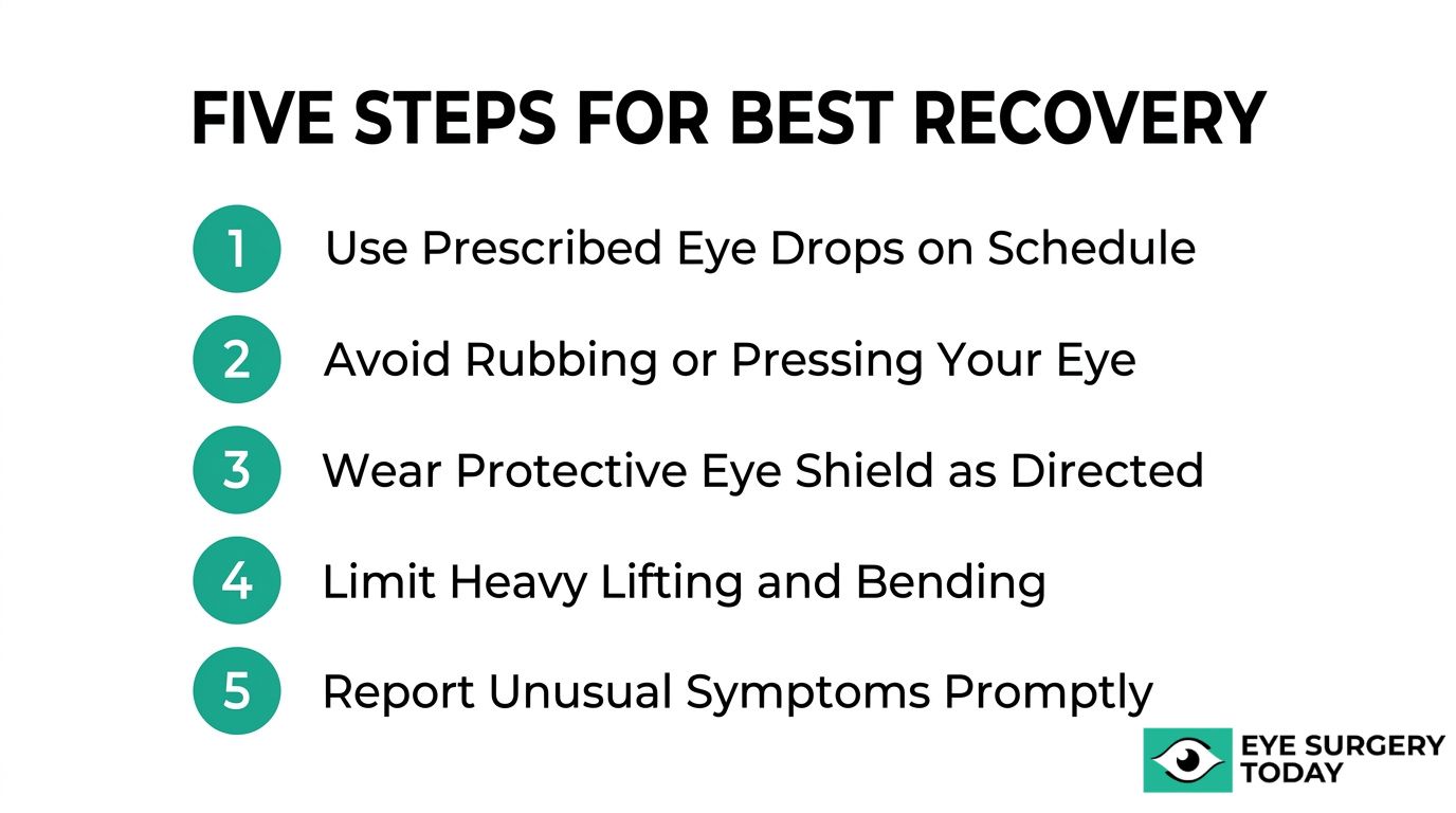
You can support the best possible vision recovery by following your surgeon’s postoperative instructions carefully, attending all scheduled follow-up appointments, and protecting your eyes during the healing period. Since full stabilization of refraction and visual acuity typically takes 4 to 8 weeks, consistent adherence to recovery protocols during this window directly influences final outcomes.
- Use prescribed eye drops on schedule. Anti-inflammatory and antibiotic drops help manage infection risk and control swelling that can temporarily blur vision.
- Avoid rubbing or pressing on the treated eye. Mechanical pressure during early healing may displace the intraocular lens or introduce bacteria.
- Wear protective eyewear as directed. A shield during sleep and sunglasses outdoors reduce exposure to dust, bright light, and accidental contact.
- Limit strenuous physical activity for the recommended period. Heavy lifting or bending can increase intraocular pressure, potentially affecting the surgical site.
- Report unusual symptoms promptly. Sudden vision loss, increasing redness, or severe pain may signal complications that require immediate attention from your eye care provider.
Dry eye symptoms deserve particular attention during recovery. According to a 2024 study published by the National Institutes of Health, nearly 20% of patients develop dry eye syndrome following cataract surgery, with 8.3% requiring nonroutine follow-up care. Using preservative-free artificial tears as recommended by your surgeon can help maintain ocular surface comfort and clarity during healing.
Managing expectations also plays an important role. While many patients notice sharper vision within the first 24 to 48 hours, subtle fluctuations in clarity, mild glare sensitivity, and occasional halos are common during the initial weeks. These symptoms typically resolve as the eye adjusts to the new intraocular lens. Patience during this stabilization period, combined with open communication with your surgical team, often makes the difference between a good recovery experience and an anxious one.
With a clear understanding of the recovery process, surgeon-reviewed educational resources can help you feel more prepared before and after your procedure.
How Can a Surgeon-Reviewed Resource Help You Prepare for Cataract Surgery?
A surgeon-reviewed resource can help you prepare for cataract surgery by providing clinically accurate, easy-to-understand information about what to expect before and after the procedure. The following sections cover how Eye Surgery Today supports informed decision-making and the essential takeaways from this guide.
Can Eye Surgery Today’s Educational Guides Help You Understand Your Vision Options?
Yes, Eye Surgery Today’s educational guides can help you understand your vision options by translating complex surgical and lens technology information into clear, patient-focused language. Each guide is surgeon-reviewed to ensure clinical accuracy without relying on confusing medical jargon.
The platform covers critical decision points, such as:
- How different intraocular lens types may affect distance, intermediate, and near vision after surgery.
- What realistic visual outcomes look like based on current clinical evidence.
- How pre-existing eye conditions or cataract severity may influence your personal results.
- What the typical recovery timeline involves, from the first 24 hours through full stabilization.
For patients weighing options between monofocal, multifocal, toric, or extended depth of focus IOLs, having access to unbiased, evidence-based comparisons is essential. Too often, patients enter consultations without a baseline understanding of what each lens can realistically deliver. Eye Surgery Today bridges that knowledge gap so patients can ask better questions and collaborate more effectively with their surgical team. This also includes specialized considerations for parents navigating infant cataract surgery success rates and outcomes for their children.
What Are the Key Takeaways About Before and After Vision Results of Cataract Surgery?
The key takeaways about before and after vision results of cataract surgery center on how significantly the procedure can restore functional vision, while setting realistic expectations for recovery.
Before surgery, cataracts progressively impair visual acuity, contrast sensitivity, color perception, and night vision. These deficits worsen as lens opacity increases, eventually interfering with daily tasks like driving and reading.
After surgery, outcomes are generally favorable. According to a large-scale study of 368,256 cataract surgeries reviewed through MDinteractive’s MIPS quality data, corrected distance visual acuity of 20/40 or better was achieved in 94.3% of cases.
Essential points to remember include:
- Vision often improves noticeably within 24 to 48 hours, but full stabilization typically takes 4 to 8 weeks.
- IOL selection directly shapes your visual outcome at different distances.
- Pre-existing conditions, cataract severity, and age may influence final results.
- Complications like posterior capsule opacification or dry eye can temporarily affect vision quality but are generally treatable.
No single lens or surgical approach guarantees a perfect outcome for every patient, which makes informed preparation one of the most valuable steps before surgery.