Cataract surgery is one of the most commonly performed procedures in ophthalmology, and while serious complications are uncommon, they can occur at any stage of the process. Understanding what may go wrong, and what to do about it, helps patients make informed decisions and respond appropriately if recovery does not follow the expected path.
This guide covers common complications and their clinical significance, warning signs and when to seek urgent care, treatment protocols for specific postoperative problems, risk factors and prevention strategies, and how surgeon experience and patient advocacy influence outcomes.
Complications range from posterior capsule opacification, the most frequently reported long-term issue, to rare but serious conditions like endophthalmitis and retained lens fragments. Each complication carries distinct symptoms, timelines, and treatment pathways that patients benefit from recognizing early.
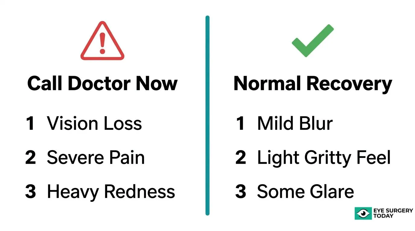
Knowing which symptoms qualify as emergencies and which can safely wait for a scheduled follow-up may prevent both unnecessary panic and dangerous delays. Sudden vision loss, severe pain, and worsening redness signal urgent evaluation, while mild grittiness and fluctuating clarity often reflect normal healing.
Treatment options vary widely depending on the complication. Some issues resolve with medication adjustments, others require laser procedures like YAG capsulotomy, and a few demand additional surgery such as IOL exchange or vitrectomy. Patients with conditions like pseudoexfoliation syndrome, diabetes, or prior eye surgery may face elevated risk profiles that influence both surgical planning and postoperative monitoring.
Surgeon selection, adherence to prescribed eye drop regimens, and consistent follow-up attendance all play measurable roles in reducing complication risk and improving visual outcomes when problems do arise.
What Are the Most Common Cataract Surgery Complications?
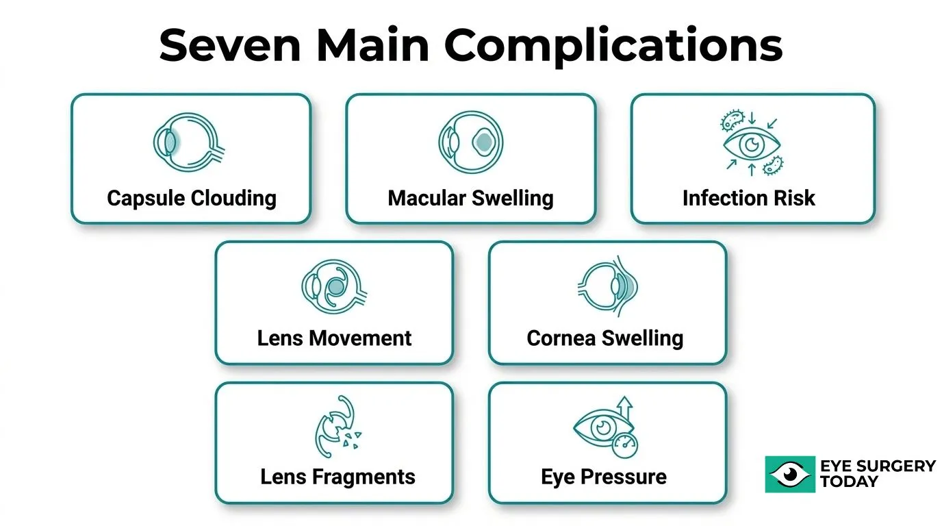
The most common cataract surgery complications include posterior capsule opacification, cystoid macular edema, endophthalmitis, intraocular lens dislocation, corneal edema, retained lens fragments, and elevated intraocular pressure. The sections below explain each complication, how it develops, and what makes it clinically significant.
What Is Posterior Capsule Opacification (PCO)?
Posterior capsule opacification (PCO) is the most common complication of cataract surgery. According to a study published in the Journal of Clinical Medicine, PCO causes a gradual deterioration of visual acuity that would otherwise improve after a successful procedure. It occurs when residual lens epithelial cells migrate across the posterior capsule, which is left in place during surgery to support the intraocular lens (IOL). Although treatable with a quick laser procedure called YAG capsulotomy, PCO can significantly affect quality of vision if left unaddressed.
What Is Cystoid Macular Edema After Cataract Surgery?
Cystoid macular edema after cataract surgery is a postoperative inflammation that causes fluid-filled cysts to form in the central retina, blurring central vision. A study published in Arq Bras Oftalmol found an overall cystoid macular edema rate of 0.82%, though the rate rose to 2.20% among patients treated with generic ketorolac 0.45% and prednisolone 1% postoperatively. Symptoms can overlap with normal post-surgical discomfort, since mild redness, grittiness, and clear discharge are expected after cataract surgery. Notably, noncompliance with prescribed eye drops remains common despite strong evidence supporting their role in preventing this complication.
What Is Endophthalmitis and How Serious Is It?
Endophthalmitis is a severe intraocular infection that represents one of the most serious complications following cataract surgery. It typically develops within days of the procedure and may cause rapid, devastating vision loss if not treated urgently. Symptoms include significant pain, pronounced redness, reduced vision, and hypopyon (pus visible in the anterior chamber). Prompt diagnosis and intravitreal antibiotic injection are essential to preserve vision. Because of its potential to cause permanent damage, any sudden worsening of vision after surgery warrants immediate evaluation.
What Is Intraocular Lens Dislocation?
Intraocular lens (IOL) dislocation is the displacement of the implanted lens from its intended position inside the eye after cataract surgery. The IOL is designed to sit within the capsular bag; if the supporting capsule or zonular fibers are weakened, the lens can shift, causing blurred or distorted vision. According to the U.S. Food and Drug Administration, phakic intraocular lenses are made of plastic or silicone and are implanted permanently to reduce dependence on glasses or contact lenses, distinguishing them from the replacement IOLs used in cataract surgery. Late dislocation, occurring months to years after surgery, is particularly associated with pre-existing conditions that compromise zonular integrity.
What Is Corneal Edema After Cataract Surgery?
Corneal edema after cataract surgery is swelling of the cornea caused by damage to or dysfunction of the endothelial cells responsible for keeping the cornea clear. Surgical trauma, ultrasound energy used during phacoemulsification, and pre-existing endothelial disease can all contribute. According to the University of Iowa Ophthalmology and Visual Sciences (EyeRounds), Fuchs’ endothelial corneal dystrophy (FECD), the most common primary corneal endothelial dystrophy, affects 4–7% of individuals over 40 and is characterized by diurnal fluctuation in visual acuity from corneal edema buildup. Patients with FECD carry a meaningfully higher risk of persistent corneal edema following cataract surgery.
What Is Retained Lens Fragment?
Retained lens fragment is a complication in which pieces of the cataract’s nucleus or cortex are not fully removed and fall into the vitreous cavity during surgery. This typically occurs when the posterior capsule ruptures, allowing fragments to drop posteriorly where standard cataract instruments cannot retrieve them. Retained fragments can trigger severe intraocular inflammation, elevated eye pressure, and retinal damage. Referral to a vitreoretinal surgeon for pars plana vitrectomy is usually required to remove the displaced material safely.
What Is Elevated Intraocular Pressure After Cataract Surgery?
Elevated intraocular pressure (IOP) after cataract surgery is an abnormal rise in eye pressure occurring in the postoperative period, which can damage the optic nerve if left untreated. A 10-year retrospective cohort study published in the Journal of Cataract and Refractive Surgery found that patients with pseudoexfoliation syndrome had significantly higher postoperative rates of elevated IOP, corneal edema, uveitis, and IOL subluxation, and were more likely to require a second operation within 90 days. Pressure spikes can result from retained viscoelastic material, inflammation, or pre-existing glaucoma, making close postoperative monitoring especially important for high-risk patients.
What Are the Warning Signs That Something Went Wrong After Cataract Surgery?
The warning signs that something went wrong after cataract surgery range from vision-threatening emergencies requiring same-day care to mild discomforts that resolve on their own. The H3 sections below cover emergency symptoms, symptoms safe to monitor, and how to distinguish normal recovery from abnormal signs.
What Symptoms Indicate a Vision-Threatening Emergency?
The symptoms that indicate a vision-threatening emergency after cataract surgery include sudden vision loss, severe eye pain, increasing redness, and significant light sensitivity. Floaters or flashes of light can signal retinal detachment, which requires immediate evaluation. According to the American Academy of Ophthalmology, if redness is accompanied by pain, light sensitivity, or a change in vision, patients should see their ophthalmologist right away. Endophthalmitis, though rare, can progress rapidly and cause irreversible damage if not treated within hours. Any single one of these symptoms after surgery warrants an urgent call to your surgical team, not a wait-and-see approach.
What Symptoms Can Wait for a Scheduled Follow-Up?
The symptoms that can wait for a scheduled follow-up include mild blurring that gradually improves, minor glare or halos around lights, and fluctuating vision during the early healing period. Slight sensitivity to bright light that diminishes day by day is generally expected in the first week. Persistent but non-worsening visual haze several weeks post-surgery may suggest posterior capsule opacification, a common late-developing complication that is treatable and non-urgent. These symptoms should still be documented and reported at your next appointment so your surgeon can assess whether any intervention is needed.
What Is Normal Discomfort Versus an Abnormal Symptom?
Normal discomfort after cataract surgery includes a gritty or itchy sensation in the eye, mild redness, and some clear watery discharge. According to Hull University Teaching Hospitals NHS Trust, these experiences are expected and typically resolve within a few days as the eye heals. Abnormal symptoms are those that worsen rather than improve, such as increasing pain, deepening redness, purulent discharge, or a sudden drop in vision. The practical distinction is trajectory: normal recovery improves steadily day over day, while abnormal signs escalate or fail to resolve. When in doubt, contacting your surgeon is always the safer choice.
How Soon Can Cataract Surgery Complications Appear?
Cataract surgery complications can appear at any stage: during the procedure itself, within the first 48 hours, or weeks to months after surgery. The sections below cover intraoperative risks, early postoperative complications, and delayed complications that develop over a longer timeline.
What Complications Can Occur During the Surgery Itself?
Complications that can occur during cataract surgery itself include posterior capsule rupture, vitreous loss, zonular dialysis, and a dropped nucleus. These intraoperative events happen while the surgeon is removing the clouded lens and positioning the intraocular lens (IOL). Patients with pseudoexfoliation syndrome face a notably higher intraoperative risk: according to a 10-year retrospective cohort study published in the Journal of Cataract and Refractive Surgery, these patients were significantly more likely to experience zonular dialysis and dropped nucleus during the procedure. Intraoperative complications matter because their severity directly influences whether postoperative problems follow. Catching anatomical risk factors before surgery, through thorough preoperative assessment, remains the most effective way to reduce these events.
What Complications Typically Emerge in the First 48 Hours?
Complications that typically emerge in the first 48 hours include elevated intraocular pressure, corneal edema, postoperative uveitis, and early signs of endophthalmitis. Some redness, grittiness, and clear discharge are expected and normal during this period. However, the American Academy of Ophthalmology states that redness accompanied by pain, light sensitivity, or a change in vision requires immediate evaluation by an ophthalmologist. Early-onset endophthalmitis, though rare, is one of the most time-sensitive complications, as delayed treatment significantly worsens outcomes. Patients should treat any sudden vision change in the first two days as urgent until proven otherwise.
What Complications Can Develop Weeks or Months Later?
Complications that can develop weeks or months after cataract surgery include posterior capsule opacification (PCO), cystoid macular edema, and IOL dislocation. PCO is the most common long-term complication; a study published in Eye found its incidence ranged from 4.7% to 18.6% at three years and 7.1% to 22.6% at five years. Cystoid macular edema, while less common, can emerge in the weeks following surgery as postoperative inflammation develops in the macula. These delayed complications reinforce why attending all scheduled follow-up appointments matters, even when early recovery feels straightforward.
What Happens If the Wrong IOL Power Is Implanted?
Implanting an IOL with the wrong power is a known, correctable complication that leaves patients with unexpected refractive error after cataract surgery. The following sections cover whether the lens can be replaced, when IOL exchange surgery is appropriate, and when a piggyback lens offers a better alternative.

Can an Incorrectly Powered IOL Be Replaced?
Yes, an incorrectly powered IOL can be replaced, though the timing and feasibility depend on several clinical factors. In the early postoperative period, the lens is easier to remove because fibrous adhesion between the IOL and surrounding tissue has not yet fully formed. Once significant scarring develops, removal becomes more technically demanding and carries greater surgical risk. According to a meta-analysis published in Diagnostics (Basel), prospective randomized studies found that 87–100% of patients using optical biometry achieved within ±1.0 D of target refraction, compared to 71–80% with applanation biometry, highlighting that biometry method influences how often power errors occur in the first place. Replacement is most straightforward when the refractive surprise is large enough that glasses or contact lenses cannot reasonably compensate.
What Is IOL Exchange Surgery and When Is It Needed?
IOL exchange surgery is a procedure in which the originally implanted intraocular lens is surgically removed and replaced with a lens of the correct power. It is typically considered when the refractive error is significant, stable, and not manageable with spectacle correction or laser enhancement. The procedure carries inherent risks, including posterior capsule damage, corneal endothelial cell loss, and increased inflammation, so surgeons generally weigh these risks against the degree of visual dissatisfaction. IOL exchange tends to be preferred when the error is large, when the original lens is still mobile, or when the patient’s anatomy makes a supplementary piggyback lens technically unsuitable.
What Is Piggyback IOL Implantation and When Is It Used?
Piggyback IOL implantation is a technique in which a second supplementary intraocular lens is inserted into the eye alongside the existing IOL to correct residual refractive error. It is used primarily when removing the original IOL carries too high a risk, such as when the lens is well-fixated within the capsular bag or when the patient’s corneal endothelium is vulnerable. The supplementary lens is typically placed in the ciliary sulcus, in front of the primary lens. This approach avoids disturbing the existing IOL-capsule complex, making it a lower-risk option for patients who would otherwise face a difficult exchange procedure. Understanding both options allows patients to have an informed conversation with their surgeon about which correction strategy best fits their individual anatomy and refractive outcome.
How Can Patients Reduce Their Risk of Cataract Surgery Complications?
Patients can reduce their risk of cataract surgery complications by preparing thoroughly before surgery, following post-operative instructions precisely, and attending all scheduled follow-up appointments. The sections below cover pre-surgical steps, critical recovery instructions, and the follow-up schedule patients should maintain.
What Should Patients Do Before Surgery to Lower Risk?
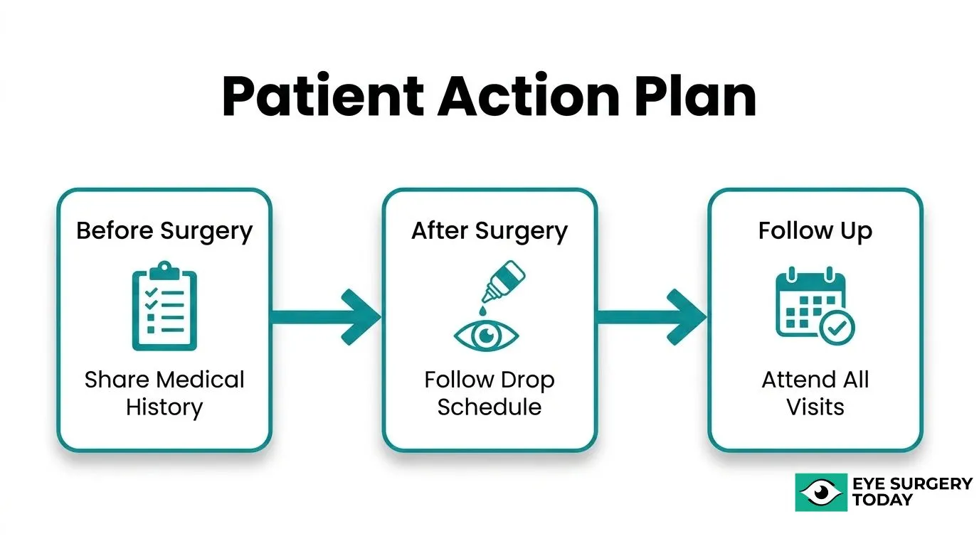
Before surgery, patients should disclose all medications, allergies, and pre-existing conditions to their surgical team. This information directly shapes antibiotic prophylaxis decisions. According to the European Society of Cataract and Refractive Surgeons (ESCRS), standard pre- and post-operative antibiotic protocols use vancomycin (1 mg) plus ceftazidime (2 mg) as the first-choice combination, with vancomycin (1 mg) plus amikacin (0.4 mg) as the second choice for patients sensitive to beta-lactam antibiotics. Reporting known drug allergies before surgery ensures the correct protocol is selected and reduces the risk of preventable infection.
What Post-Operative Instructions Are Most Critical to Follow?
The most critical post-operative instructions are using prescribed eye drops as directed, avoiding water exposure to the eye, refraining from rubbing the eye, and attending all follow-up appointments. Noncompliance with prescribed eye drops remains a common issue despite strong evidence supporting their role in preventing complications such as cystoid macular edema and infection. Patients should also avoid strenuous activity and wear any protective eye shield recommended by their surgeon during the early recovery period.
What Follow-Up Schedule Should Patients Maintain After Surgery?
Patients should maintain a follow-up schedule that typically includes visits at 24 hours, one week, and four to six weeks after surgery, with additional visits if new symptoms arise. Consistent follow-up allows clinicians to catch complications before they affect long-term vision. A study published in Translational Vision Science and Technology found that 40% of participants still had vision impairment (BCVA less than 6/18 Snellen acuity) following cataract surgery treatment, reinforcing that outcomes are not uniform and that active monitoring is essential. Skipping follow-up visits is one of the most underestimated risks patients take after an otherwise successful procedure.
How Does Surgeon Experience Affect Cataract Surgery Outcomes?
Surgeon experience affects cataract surgery outcomes through complication rates, procedural range, and overall visual results. The sections below cover how surgeon volume and training background influence patient outcomes and what to ask when evaluating a surgeon.
Does a More Experienced Surgeon Produce Better Visual Results?
A more experienced surgeon does not always produce measurably better visual acuity outcomes in routine cases. A study published in Ophthalmology Science analyzed 14,537 cataract surgery cases over 32 months and found no statistically significant difference in the proportion of cases achieving better postoperative visual acuity between resident and attending surgeons. Among 337 resident-performed surgeries, 74% achieved better postoperative visual acuity and 51% showed more than two lines of improvement.
That said, visual acuity alone does not capture the full picture. Complex cases, uncommon anatomical variants, and patients with pre-existing ocular conditions may genuinely benefit from a surgeon with greater procedural depth and subspecialty training.
What Complication Rate Should You Expect From an Experienced Surgeon?
The complication rate you should expect from an experienced cataract surgeon is well below 5% overall, with major complications occurring at approximately 1 in 2,000 cases, according to University of Utah Health. Asking a surgeon directly for their outcome statistics is one of the most practical ways to evaluate their track record before surgery.
Fellowship-trained surgeons tend to perform a broader variety of procedures, which may be relevant when patients present with higher-risk profiles or complex anatomy requiring intraoperative judgment beyond routine phacoemulsification.
What Questions Should You Ask Your Surgeon If Something Feels Wrong?
The questions you should ask your surgeon if something feels wrong cover your symptoms, the next diagnostic steps, treatment options, and realistic outcome expectations. Knowing which questions to raise helps you advocate effectively and ensures nothing gets dismissed as routine discomfort when it warrants closer attention.
What Should You Ask About Your Specific Symptoms?
The first questions to ask about your specific symptoms are whether what you are experiencing is expected at this stage of recovery and how it compares to what your surgeon typically sees in patients at the same point post-operatively. Ask your surgeon to describe exactly which features of your symptom, such as the location, severity, or timing, concern them most. Request clarification on whether the symptom could indicate a treatable complication like cystoid macular edema or elevated intraocular pressure, or whether it is consistent with normal healing. According to the American Academy of Ophthalmology, redness accompanied by pain, light sensitivity, or a change in vision warrants immediate evaluation rather than a watchful waiting approach.
What Should You Ask About Next Steps and Testing?
The questions to ask about next steps and testing include what specific examination or imaging your surgeon plans to perform, how quickly results will be available, and what the findings will determine about your care. Ask whether a slit-lamp examination, optical coherence tomography, or intraocular pressure measurement is indicated. It is also reasonable to ask what threshold finding would prompt a referral to a specialist or a change in your current treatment plan. Surgeons who welcome these questions and answer them concisely are demonstrating the transparency that experienced, high-volume practices tend to prioritize.
What Should You Ask About Treatment Options and Timelines?
The questions to ask about treatment options and timelines are what interventions are available, which is recommended first, and how soon you should expect improvement. Ask whether the proposed treatment, such as additional eye drops, a laser procedure, or a surgical revision, addresses the underlying cause or only manages symptoms. Clarify what the monitoring schedule will look like if a wait-and-see approach is chosen, and at what point the plan would change. Understanding the timeline for expected improvement gives you a concrete benchmark, making it far easier to recognize if your recovery is genuinely stalling rather than simply progressing more slowly than hoped.