Glaucoma is a group of eye conditions that damage the optic nerve through progressive loss of retinal ganglion cells, often without noticeable symptoms until significant vision loss has already occurred.
This guide covers who should be screened and why, the specific diagnostic tests used to evaluate glaucoma, how to interpret your results, and what happens after a diagnosis is confirmed.
Certain populations face substantially higher glaucoma risk. Age, ethnicity, family history, elevated intraocular pressure, and systemic conditions like obstructive sleep apnea all influence when screening should begin and how frequently it should occur.
A comprehensive glaucoma evaluation relies on multiple complementary tests rather than any single measurement. Tonometry assesses eye pressure, ophthalmoscopy and OCT examine optic nerve structure, perimetry maps functional vision loss, and gonioscopy classifies the type of glaucoma present. Each test captures a different dimension of the disease.
Interpreting results requires clinical context because as many as one in five test results may be inaccurate due to false positives or false negatives. Corneal thickness, patient fatigue, and media opacities can all affect reliability, which is why ophthalmologists typically confirm findings across multiple tests before reaching conclusions.
Once glaucoma is confirmed, treatment focuses on lowering intraocular pressure through medications, laser procedures, or surgery, with follow-up intervals tailored to disease severity. Consistent monitoring remains one of the most impactful steps for preserving vision over time.
What Is Glaucoma and Why Does Early Diagnosis Matter?
Glaucoma is a group of eye conditions that damage the optic nerve, and early diagnosis matters because vision lost to glaucoma cannot be restored. The following sub-sections define the condition, explain its main types, and outline why catching it before symptoms appear is critical.
What Is Glaucoma?
Glaucoma is an acquired loss of retinal ganglion cells and axons within the optic nerve that results in a characteristic optic nerve head appearance and progressive vision loss, according to StatPearls via the National Center for Biotechnology Information. Aqueous humor, the fluid produced by the ciliary body, drains through the trabecular meshwork into Schlemm canal. When this drainage system impaired, intraocular pressure (IOP) can rise and contribute to optic nerve damage. Because the process is typically painless and slow, patients rarely notice changes until significant damage has already occurred.
What Are the Main Types of Glaucoma?
The main types of glaucoma are open-angle glaucoma, angle-closure glaucoma, normal-tension glaucoma, and secondary forms such as neovascular and uveitic glaucoma. Each type differs in cause and onset:
- Primary open-angle glaucoma (POAG): The most common type, affecting 9 in 10 Americans with glaucoma, involving slow, painless optic nerve damage from impaired aqueous drainage at the trabecular meshwork.
- Acute angle-closure glaucoma: A sudden blockage of the drainage angle by the iris, causing rapid vision decline, eye pain, headache, nausea, and corneal edema.
- Normal-tension glaucoma: Optic nerve damage that progresses despite IOP readings consistently below 21 mmHg.
- Neovascular glaucoma: Caused by abnormal new blood vessels covering the drainage area, typically linked to diabetes or high blood pressure.
- Uveitic glaucoma: Develops in approximately 2 in 10 people with uveitis, where inflammation and scar tissue block fluid drainage.
Why Does Early Detection of Glaucoma Matter?
Early detection of glaucoma matters because the disease destroys optic nerve fibers silently, and any vision lost before diagnosis is permanent. More than 3 million Americans currently have glaucoma, according to the CDC, with projections estimate that more than 7.3 million people in the U.S. will have open-angle glaucoma by 2050. Open-angle glaucoma, the dominant form, produces no pain or early visual symptoms, so most patients are unaware of their condition until substantial nerve damage has already occurred. Routine screening allows clinicians to identify elevated IOP, optic nerve changes, or retinal nerve fiber thinning before functional vision loss begins, making intervention possible when treatment is most effective.
Who Should Get Tested for Glaucoma?
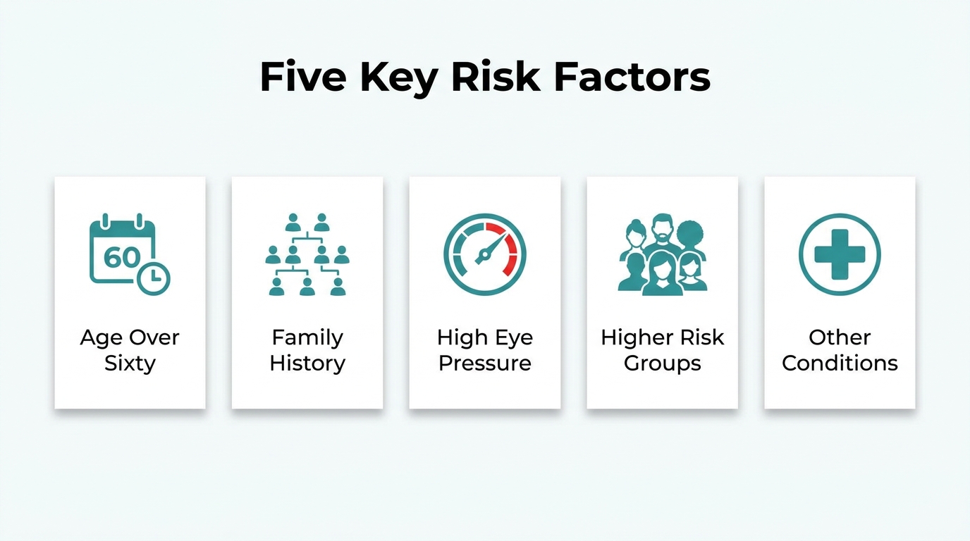
People at higher risk for glaucoma should get tested before symptoms appear. Key risk factors include age, family history, elevated eye pressure, ethnicity, and certain medical conditions.
Who Is at Higher Risk Due to Age?
People at higher risk due to age are those over 60, as glaucoma prevalence rises significantly with each decade of life. Among individuals aged 40 and older, the prevalence of glaucoma is 2.56%, according to a study reported by Ophthalmology Times. Risk accelerates further after age 60, making routine screening increasingly important as patients grow older. For adults over 40 with no other risk factors, regular comprehensive eye exams remain the most reliable way to catch early optic nerve changes before vision loss occurs.
Who Is at Higher Risk Due to Family History?
People at higher risk due to family history are those with a first-degree relative diagnosed with glaucoma. Genetic mutations contribute meaningfully to disease risk; mutations in the myocilin (MYOC) gene are present in 2 to 4% of primary open-angle glaucoma (POAG) patients, according to a review published in PMC. Because glaucoma often progresses without symptoms, a family history should prompt earlier and more frequent screening, even when eye pressure appears normal.
Who Is at Higher Risk Due to Elevated Eye Pressure?
People at higher risk due to elevated eye pressure are those with consistently high intraocular pressure (IOP), a primary modifiable risk factor for glaucomatous optic nerve damage. The relationship between blood pressure and IOP is nuanced: patients with very high blood pressure tend to have higher IOP, while those with very low blood pressure face an increased risk of open-angle glaucoma, thought to be related to poor blood flow to the optic nerve, according to the BrightFocus Foundation. For glaucoma suspects with stable ocular findings, the American Academy of Ophthalmology (AAO) recommends follow-up intervals of 6 to 12 months, depending on the individual’s IOP levels and overall risk profile. Elevated IOP alone does not confirm glaucoma, but it warrants structured monitoring to detect any progression early.
Who Is at Higher Risk Due to Ethnicity?
People at higher risk due to ethnicity include African Americans, Latinos, and Asian Americans, each facing distinct patterns of glaucoma risk. The Baltimore Eye Survey found that glaucoma prevalence in individuals of African descent was six times that of European Americans in some age groups, with the condition also diagnosed approximately 10 years earlier and progressing more rapidly. In the Los Angeles Latino Eye Study (LALES), open-angle glaucoma prevalence among Latinos of primarily Mexican ancestry was approximately 5%, comparable to rates in African Americans. Open-angle glaucoma prevalence in Asian Americans was 6.52%, higher than the 5.59% observed in non-Hispanic whites, according to data published in ScienceDirect. These disparities make ethnicity an important factor when determining the appropriate age to begin and frequency of glaucoma screening.
Who Is at Higher Risk Due to Other Medical Conditions?
People at higher risk due to other medical conditions include those with obstructive sleep apnea (OSA), diabetes, and blood pressure disorders. Glaucoma prevalence was found to be 16% among individuals with OSA, compared to 8% in those without the condition, according to a study published in PMC. Diabetes is associated with neovascular glaucoma, a form caused when abnormal blood vessels cover the eye’s drainage area. These systemic conditions share mechanisms, such as impaired blood flow and elevated vascular resistance, that may compromise optic nerve health. Patients managing these conditions should inform their eye care provider, as earlier and more frequent testing may be warranted.
What Are the Main Tests Used to Diagnose Glaucoma?
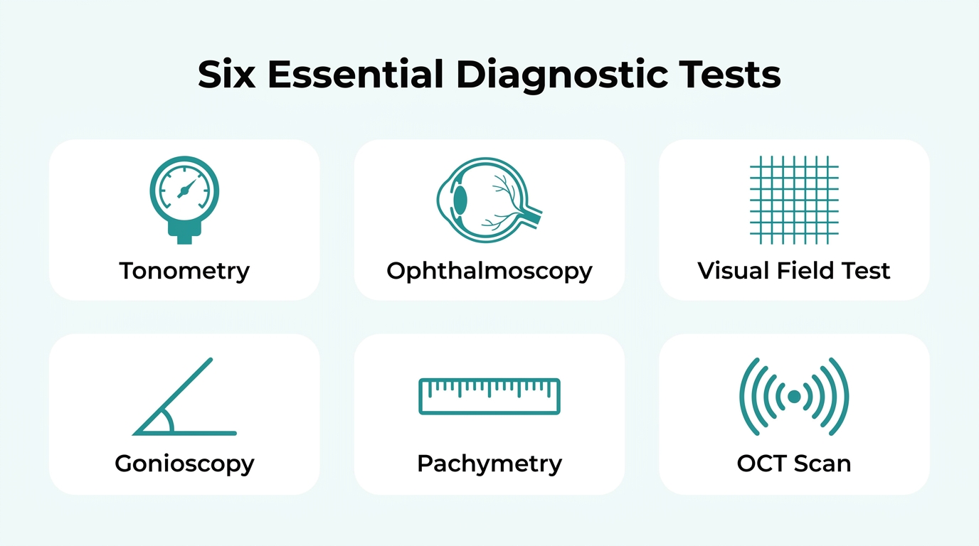
The main tests used to diagnose glaucoma are tonometry, ophthalmoscopy, perimetry, gonioscopy, pachymetry, and optical coherence tomography (OCT). Each test examines a different aspect of eye health, and together they give clinicians a complete picture of glaucomatous damage and disease risk.
Tonometry
Tonometry measures intraocular pressure (IOP), one of the primary indicators of glaucoma risk. A clinician applies a small instrument or air pulse to the cornea to assess how much resistance the eye exerts. Normal IOP typically falls between 10 and 21 mmHg, and readings above this range may prompt further evaluation. Because corneal thickness can affect pressure readings, tonometry results are often interpreted alongside pachymetry data for accuracy.
Ophthalmoscopy
Ophthalmoscopy examines the optic nerve for structural signs of glaucomatous damage. A dilated eye exam uses drops to open the pupil, allowing the clinician to shine bright light and magnify the retina and optic nerve directly, according to Cleveland Clinic. A vertical cup-to-disc (C/D) ratio of 0.6 or greater is often used as a threshold for suspected glaucomatous damage, based on the 97.5th percentile of the C/D ratio distribution in healthy eyes. This makes ophthalmoscopy essential for catching structural changes before vision loss occurs.
Perimetry (Visual Field Test)
Perimetry maps the patient’s field of vision to detect areas of sight loss caused by glaucoma. Automated static perimetry requires the patient to look into a machine and press a button when lights appear, identifying whether peripheral or central vision has been lost. Glaucomatous damage typically appears first in the peripheral field, making this test particularly valuable for detecting early and mid-stage disease progression.
Gonioscopy
Gonioscopy evaluates the drainage angle between the cornea and iris to classify glaucoma type. During the procedure, an ophthalmologist uses eye drops to numb the eye and places a special mirrored lens against the cornea to directly visualize whether the drainage angle is open or closed, as described by Cleveland Clinic. This distinction is clinically critical: open-angle and closed-angle glaucoma follow different treatment pathways, and an incorrect classification can lead to ineffective management.
Pachymetry
Pachymetry measures central corneal thickness (CCT), a key variable in interpreting IOP readings accurately. The procedure involves numbing the eye with drops and touching the cornea with a small probe, according to Cleveland Clinic. Thinner corneas tend to produce artificially low tonometry readings, while thicker corneas can produce falsely elevated ones. This is especially relevant for patients who have undergone refractive corneal procedures, where tonometric accuracy is further complicated.
Optical Coherence Tomography (OCT)
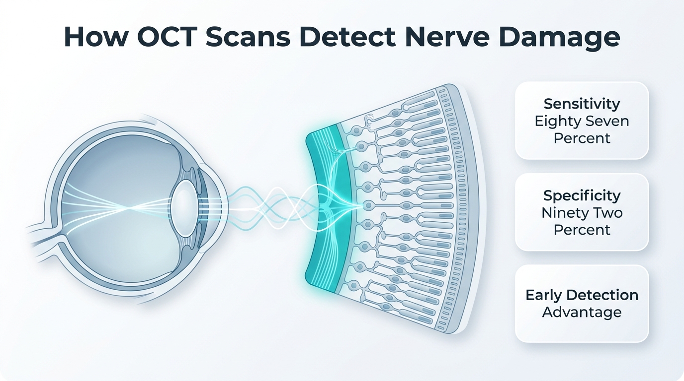
Optical coherence tomography (OCT) produces detailed cross-sectional images of the retinal nerve fiber layer (RNFL) to detect glaucomatous thinning. The procedure involves the patient resting their chin on a machine while a digital camera captures images of each layer of eye tissue, as described by Cleveland Clinic. According to Eyes On Eyecare, OCT RNFL thickness measurements achieve a combined sensitivity of 87% and specificity of 92% for glaucoma diagnosis, making it one of the most precise structural tools available. Given these metrics, OCT can help detect structural nerve changes that may appear before functional vision loss becomes measurable on perimetry.
With each of these tests covering a distinct diagnostic dimension, a complete glaucoma evaluation rarely relies on any single result alone.
How Does Tonometry Measure Eye Pressure?
Tonometry measures eye pressure by assessing the force required to flatten or indent the corneal surface, which correlates directly with intraocular pressure (IOP). The main methods vary by technique, accuracy, and clinical setting, with corneal properties playing a central role in result reliability.
Goldmann Applanation Tonometry
Goldmann applanation tonometry (GAT) is the gold standard for measuring intraocular pressure in clinical practice. According to Medscape’s Primary Open-Angle Glaucoma Workup, GAT accuracy depends on corneal rigidity, curvature, and central corneal thickness (CCT), meaning readings can be falsely elevated or underestimated in eyes with atypical corneal profiles. This makes pachymetry a critical companion test: by numbing the eye and touching the cornea with a small probe, clinicians measure CCT and adjust tonometry readings accordingly. Eyes with prior refractive surgery are particularly susceptible to tonometric error, making this correction especially important.
Non-Contact and Rebound Tonometry
Non-contact tonometry uses a rapid air impulse to flatten the cornea without touching the eye, making it useful for screening. The Ocular Response Analyzer (ORA) builds on this by recording two applanation pressure measurements with a rapid air impulse to account for corneal biomechanical properties, offering a more refined IOP estimate. The iCare rebound tonometer offers a touch-based alternative that requires no anesthetic. However, according to EyeWiki’s review of IOP measurement reliability, the iCare rebound tonometer demonstrates only 73% concordance within 5 mmHg of GAT measurements, suggesting it is best suited for screening rather than definitive clinical assessment.
What Does a Visual Field Test Reveal About Glaucoma?
A visual field test reveals the presence, location, and severity of peripheral and central vision loss caused by glaucomatous optic nerve damage. By mapping areas where light sensitivity is reduced, it gives clinicians a direct measure of functional vision loss over time.
Standard automated perimetry (SAP) using the Humphrey Field Analyzer is the current standard for evaluating the visual field in glaucoma. The Humphrey Field Analyzer incorporates the Glaucoma Hemifield Test (GHT), which compares sensitivity between the upper and lower hemifields to identify asymmetric losses characteristic of glaucomatous damage, according to StatPearls published by the National Center for Biotechnology Information.
During the test, the patient looks into a machine and responds to light stimuli that appear across their field of view. The device records which points are seen and which are missed, generating a sensitivity map that highlights defects corresponding to areas of optic nerve damage.
Because single test results can be affected by patient fatigue or inattention, repeated testing strengthens diagnostic confidence. The American Academy of Ophthalmology recommends visual field testing at least once per year for patients with confirmed glaucoma to monitor for disease progression.
Visual field testing is most valuable when interpreted alongside OCT structural data, since functional loss detected by perimetry often follows structural nerve fiber layer thinning.
How Does OCT Help Detect Glaucoma Early?
OCT helps detect glaucoma early by imaging the retinal nerve fiber layer (RNFL) to identify structural damage before significant vision loss occurs. The sections below explain how OCT measures RNFL thickness and how it compares to other detection technologies.
How Does OCT Measure Retinal Nerve Fiber Layer Thickness?
OCT measures retinal nerve fiber layer thickness by using light waves to capture high-resolution cross-sectional images of each tissue layer in the eye. During the scan, the patient rests their chin on the machine while a digital camera maps the RNFL across multiple quadrants. According to a report published by Eyes On Eyecare, OCT RNFL thickness measurements achieve a combined sensitivity of 87% and specificity of 92% for glaucoma diagnosis, making it one of the more precise structural tests available. Because RNFL thinning can precede detectable visual field loss by years, OCT offers a meaningful window for early intervention.
How Does OCT Compare to Frequency-Doubling Technology for Glaucoma Detection?
OCT compares favorably to frequency-doubling technology (FDT) for glaucoma detection, particularly in earlier disease stages. FDT is a functional test that assesses contrast sensitivity, whereas OCT measures structural change directly. For detection of moderate or worse glaucoma, a study published in Ophthalmology Glaucoma found that FDT achieved a sensitivity of only 55% at a fixed specificity of 90%, a notably lower detection rate than OCT’s structural imaging approach. In clinical practice, structural OCT measurements tend to identify nerve damage at an earlier point in the disease course than functional tests like FDT, making OCT a tool for glaucoma surveillance.
What Happens During a Comprehensive Glaucoma Eye Exam?
A comprehensive glaucoma eye exam follows a structured sequence of tests covering preparation, pupil dilation, and imaging. The sections below explain what to do before arriving, what occurs once dilation begins, and how much time the full evaluation requires.
What Should You Do to Prepare Before the Exam?
Preparing before the exam involves a few practical steps to help the visit run smoothly and ensure accurate results.
- Arrange a driver. Dilation drops blur near vision for several hours, making it unsafe to drive immediately after the appointment.
- Bring a list of current medications. Certain drugs, including corticosteroids and antihistamines, can affect intraocular pressure readings.
- Bring your current glasses or contact lens prescription. This helps the examiner assess baseline visual acuity accurately.
- Remove contact lenses before tonometry. Contacts can interfere with pressure measurements; ask your provider how far in advance to remove them.
- Note any symptoms. Eye pain, halos around lights, or recent vision changes are all relevant for the examining clinician to know.
Arriving well-rested and informed allows the care team to move efficiently through each diagnostic step.
What Occurs During the Dilated Eye Examination?
The dilated eye examination uses eye drops to open the pupil, allowing the clinician to use a specialized lens with bright light to magnify the retina and optic nerve for signs of glaucomatous damage, according to the Cleveland Clinic. Once the drops take effect, typically within 15 to 30 minutes, the examiner assesses the optic nerve head for cup-to-disc changes, evaluates the retinal nerve fiber layer, and may perform optical coherence tomography imaging with the patient resting their chin on the machine while a digital camera captures cross-sectional tissue layers. Gonioscopy may also be performed at this stage, using a contact lens to visualize the drainage angle between the cornea and iris. Together, these steps provide the structural and functional data needed to characterize any nerve damage present.
How Long Does a Full Glaucoma Evaluation Take?
A full glaucoma evaluation can take longer than a routine eye exam when multiple diagnostic components are included. The time accounts for dilation onset, tonometry, visual field testing, pachymetry, OCT imaging, and the clinician’s review of findings. Visual field testing alone requires sustained focus and typically runs 15 to 20 minutes per eye. Patients should plan for extended wait times if dilation is slower or if additional imaging is needed. For context, research published in Ophthalmology Glaucoma underscores that diagnostic accuracy improves when multiple test modalities are completed in a single visit rather than spread across appointments, reinforcing the value of allowing adequate time for a thorough evaluation.
What Do Your Glaucoma Test Results Mean?
Glaucoma test results require careful interpretation because, according to a review published in Review of Ophthalmology, as many as one in five results may incorrectly indicate the presence or absence of the disease due to false positives or false negatives. The following H3 sections cover what specific result combinations typically mean, from normal pressure with suspicious discs to optic nerve thinning on OCT.
What Does Normal Eye Pressure With Suspicious Discs Mean?
Normal eye pressure with suspicious discs means the optic nerve shows structural changes consistent with glaucoma despite intraocular pressure falling within the accepted normal range. This pattern is closely associated with normal-tension glaucoma, where optic nerve damage occurs even without elevated IOP.
A cup-to-disc ratio appearing large or asymmetric is one key indicator. According to a PMC study on large disc cupping with normal IOP, a vertical cup-to-disc ratio of 0.6 or greater meets the threshold for suspected glaucomatous damage. In this scenario, an ophthalmologist will often review mean deviation (MD) values from automated perimetry, where more negative values signal greater vision loss. A reasonable initial management goal may involve reducing IOP by 20% to 30% below baseline, even when pressure appears normal.
What Does Elevated Eye Pressure With Normal Visual Fields Mean?
Elevated eye pressure with normal visual fields means the patient may have ocular hypertension, a condition that carries glaucoma risk without yet producing measurable field loss. This is an important distinction because structural damage to the optic nerve can precede detectable visual field defects by years.
Gonioscopy may be performed to determine whether the drainage angle is open or closed, as angle anatomy directly influences treatment planning. Confirmatory testing is also advised: according to a PubMed study on Glaucoma Hemifield Test repeatability, requiring two abnormal visual field results rather than one improved testing specificity from 80.8% to 89.9%. This approach reduces the risk of acting on a single unreliable result and is worth discussing with a treating ophthalmologist.
What Do Abnormal Visual Field Results Indicate?
Abnormal visual field results indicate that the patient has measurable loss of light sensitivity in specific areas of their visual field, which may correspond to glaucomatous optic nerve damage. According to StatPearls via the Humphrey Visual Field entry, the Humphrey Field Analyzer uses the Glaucoma Hemifield Test (GHT) to compare sensitivity between the upper and lower hemifields, flagging asymmetry that warrants further investigation.
Key patterns clinicians look for include:
- Arcuate scotomas: Curved blind spots following the nerve fiber layer path, typical of early-to-moderate glaucoma.
- Nasal step defects: Asymmetric loss respecting the horizontal midline, a common early finding.
- Central field loss: Present in advanced disease, indicating significant retinal ganglion cell damage.
- Mean deviation (MD) value: More negative MD scores reflect greater average sensitivity loss compared to an age-matched population.
A single abnormal result is rarely definitive on its own, and repeat testing is standard practice before treatment decisions are made.
What Does Optic Nerve Thinning on OCT Suggest?
Optic nerve thinning on OCT suggests loss of retinal ganglion cells and axons within the optic nerve, which is the defining structural hallmark of glaucoma. According to StatPearls, glaucoma is an acquired optic neuropathy where this cellular loss produces a characteristic optic nerve head appearance alongside progressive vision loss.
A diagnosis of glaucoma suspect may be established when OCT reveals circumpapillary RNFL thinning, even without elevated IOP or visual field loss, according to the American Academy of Ophthalmology’s 2025 Primary Open-Angle Glaucoma Suspect Preferred Practice Pattern. RNFL thinning typically follows the ISNT rule in healthy eyes, where the inferior quadrant is thickest; violations of this pattern can indicate glaucomatous damage. OCT findings alone rarely confirm a diagnosis but, combined with clinical exam data, they provide a strong structural basis for monitoring or initiating treatment.
How Often Should You Be Retested for Glaucoma?
Retest frequency for glaucoma depends on your diagnosis stage, risk profile, and whether intraocular pressure is controlled. The H3 sections below cover intervals for glaucoma suspects, mild glaucoma, and moderate to severe glaucoma.
How Often Should Glaucoma Suspects Be Retested?
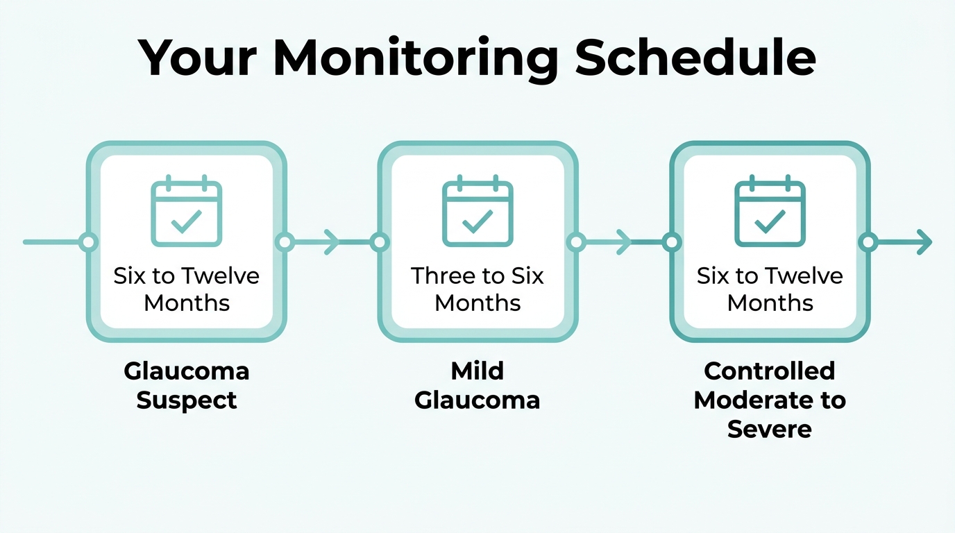
Glaucoma suspects should be retested every 6 to 12 months, depending on their individual risk profile and intraocular pressure levels. According to the American Academy of Ophthalmology’s 2025 Preferred Practice Pattern, this interval applies to suspects with stable ocular findings. Patients with higher-risk features, such as borderline IOP or suspicious optic nerve appearance, generally warrant follow-up closer to the 6-month end of that range. Consistent monitoring at these intervals allows clinicians to detect early structural or functional changes before confirmed disease develops.
How Often Should Mild Glaucoma Be Monitored?
Mild glaucoma requires follow-up approximately every 3 to 6 months when target IOP has been achieved and no progression is observed. The American Academy of Ophthalmology’s 2025 Glaucoma Summary Benchmarks establish this interval as the standard for stable, well-controlled mild disease. More frequent visits may be warranted in the first year after diagnosis to confirm that treatment is maintaining adequate pressure reduction.
How Often Should Moderate to Severe Glaucoma Be Monitored?
Moderate to severe glaucoma requires follow-up every 6 to 12 months once target IOP is achieved and no progression is noted, provided control has been maintained for more than 6 months. The AAO’s 2025 Glaucoma Summary Benchmarks specify this interval for stable advanced disease. Patients with ongoing progression or recent treatment changes typically need more frequent evaluation. In clinical practice, regular visual field testing remains important, making consistent adherence to scheduled exams one of the most impactful steps a patient can take.
What Are the Possible Risks or Limitations of Glaucoma Tests?
The possible risks or limitations of glaucoma tests include false positive results, false negative results, measurement inaccuracies caused by corneal properties, and patient-related factors that reduce reliability. Understanding these limitations helps patients and clinicians interpret results within the broader clinical picture rather than relying on any single test.
Can Glaucoma Tests Produce False Positives or False Negatives?
Yes, glaucoma tests can produce false positives or false negatives. According to a report published in Review of Ophthalmology, as many as one in five glaucoma test results may incorrectly indicate the presence or absence of the disease. A false positive may lead to unnecessary treatment, while a false negative can delay a diagnosis that could have preserved vision. This is why ophthalmologists typically confirm findings across multiple tests before reaching a definitive conclusion.
Does Corneal Thickness Affect the Accuracy of Pressure Measurements?
Corneal thickness affects the accuracy of intraocular pressure measurements directly. Goldmann applanation tonometry, the gold standard for measuring eye pressure, depends on corneal rigidity, curvature, and central corneal thickness. Thicker corneas tend to produce artificially elevated readings, while thinner corneas may underestimate true pressure. In patients who have undergone refractive corneal procedures such as LASIK, this inaccuracy is compounded, making corneal thickness measurement an essential step in any reliable pressure assessment.
Are Visual Field Tests Always Reliable for Detecting Glaucoma?
Visual field tests are not always reliable on their own for detecting glaucoma. Patient concentration, fatigue, and learning effects can all influence results, introducing variability between testing sessions. Specificity in automated visual field testing improves from 80.8% to 89.9% when two abnormal results are required rather than one, according to a study published in PubMed, though this comes with a modest reduction in sensitivity. Repeating abnormal results before acting on them is a well-established clinical practice for this reason.
What Other Factors Can Limit Glaucoma Test Results?
Several additional factors can limit glaucoma test results, including media opacities such as cataracts, pupil dilation quality, and OCT signal strength. Cataracts can reduce image clarity on OCT scans, potentially affecting how accurately retinal nerve fiber layer thickness is measured. Patient cooperation and fixation stability also influence the quality of imaging and perimetry data. These variables reinforce why a comprehensive evaluation, combining multiple tests interpreted together, remains the most dependable approach for glaucoma diagnosis.
What Happens After a Glaucoma Diagnosis Is Confirmed?
After a glaucoma diagnosis is confirmed, your ophthalmologist will establish a treatment plan focused on lowering intraocular pressure to slow disease progression, and schedule ongoing monitoring to track any changes over time.
Treatment options and follow-up schedules vary based on disease severity, and your care plan will typically address three areas: medication management, possible laser or surgical intervention, and regular retesting intervals.
What Treatment Options Are Available After Diagnosis?
Treatment options after a glaucoma diagnosis include prescription eye drops, laser procedures such as Selective Laser Trabeculoplasty (SLT), minimally invasive glaucoma surgery (MIGS), and incisional procedures like trabeculectomy. The choice depends on disease stage and how well IOP responds to initial therapy.
How Will Your Doctor Monitor Glaucoma Over Time?
Your doctor will monitor glaucoma through scheduled follow-up visits that include IOP measurement, visual field testing, and OCT imaging to detect any progression. The frequency of these visits depends on how stable your condition is and whether your target IOP has been achieved.
Are There Lifestyle or Health Factors to Address After Diagnosis?
Yes, there are health conditions that may influence glaucoma progression and should be addressed after diagnosis. According to data published by Glaucoma Australia, incident glaucoma is 33% to 43% more likely in individuals with sleep apnea compared to controls, even after adjusting for comorbidities. Managing systemic conditions such as obstructive sleep apnea may therefore be a relevant part of your overall care plan.
How Can Eye Care Resource Help You?
An eye care resource can help you by translating complex clinical findings into clear, actionable information. Eye Surgery Today covers what glaucoma tests measure, how results are interpreted, and what next steps typically look like.
Can Eye Surgery Today Help You Understand Your Options?
Eye Surgery Today can help you understand your options by providing educational content on glaucoma diagnosis, testing procedures, and treatment pathways. Glaucoma is a condition where early detection directly affects outcomes, yet many patients arrive at appointments without knowing what tests to expect or what results mean. Eye Surgery Today helps explain these topics in clear language, so you can ask better questions during your eye care visit.
What Are the Key Takeaways About Glaucoma Diagnosis and Testing?
The key takeaways about glaucoma diagnosis and testing are that early detection relies on multiple complementary tests, results require careful interpretation, and ongoing monitoring is essential. No single test confirms glaucoma alone. According to a Review of Ophthalmology analysis, as many as one in five glaucoma test results may incorrectly indicate the presence or absence of disease, underscoring why clinicians combine tonometry, visual field testing, OCT, and optic nerve imaging before drawing conclusions. Certain populations, including those with uveitis, face compounded risk since approximately 2 in 10 people with uveitis may develop uveitic glaucoma. Understanding these nuances helps you engage more confidently with your diagnosis and monitoring plan.