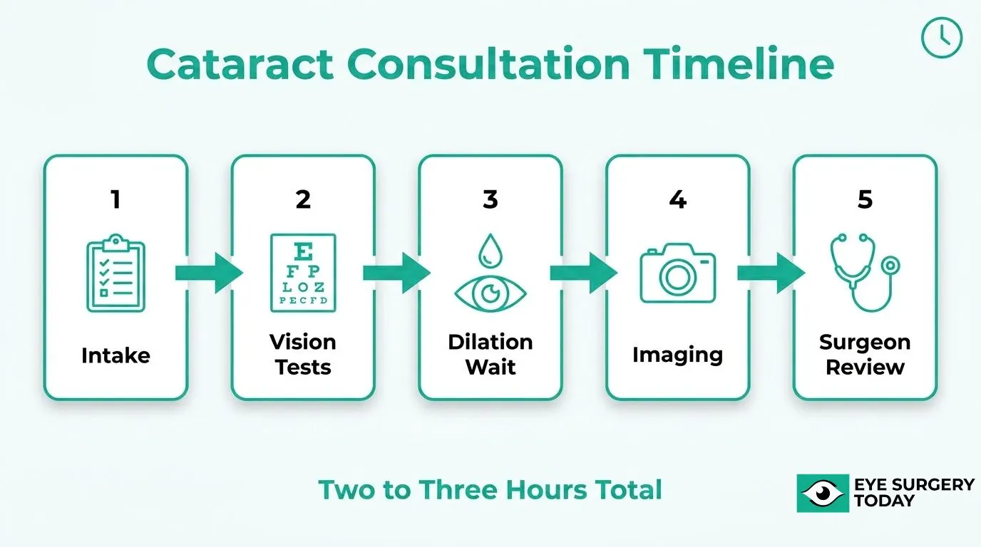
A cataract consultation is a pre-operative appointment where a specialist team examines, measures, and maps your eye to determine surgical suitability and select the correct intraocular lens. The visit typically requires 2 to 3 hours because it combines multiple clinical stages that must occur in sequence.
This guide covers the consultation workflow and what happens at each stage, why eye dilation adds significant waiting time, the diagnostic tests and measurements performed, how the right lens is chosen, who you will meet during the visit, how to prepare, and what happens between the consultation and your surgery date.
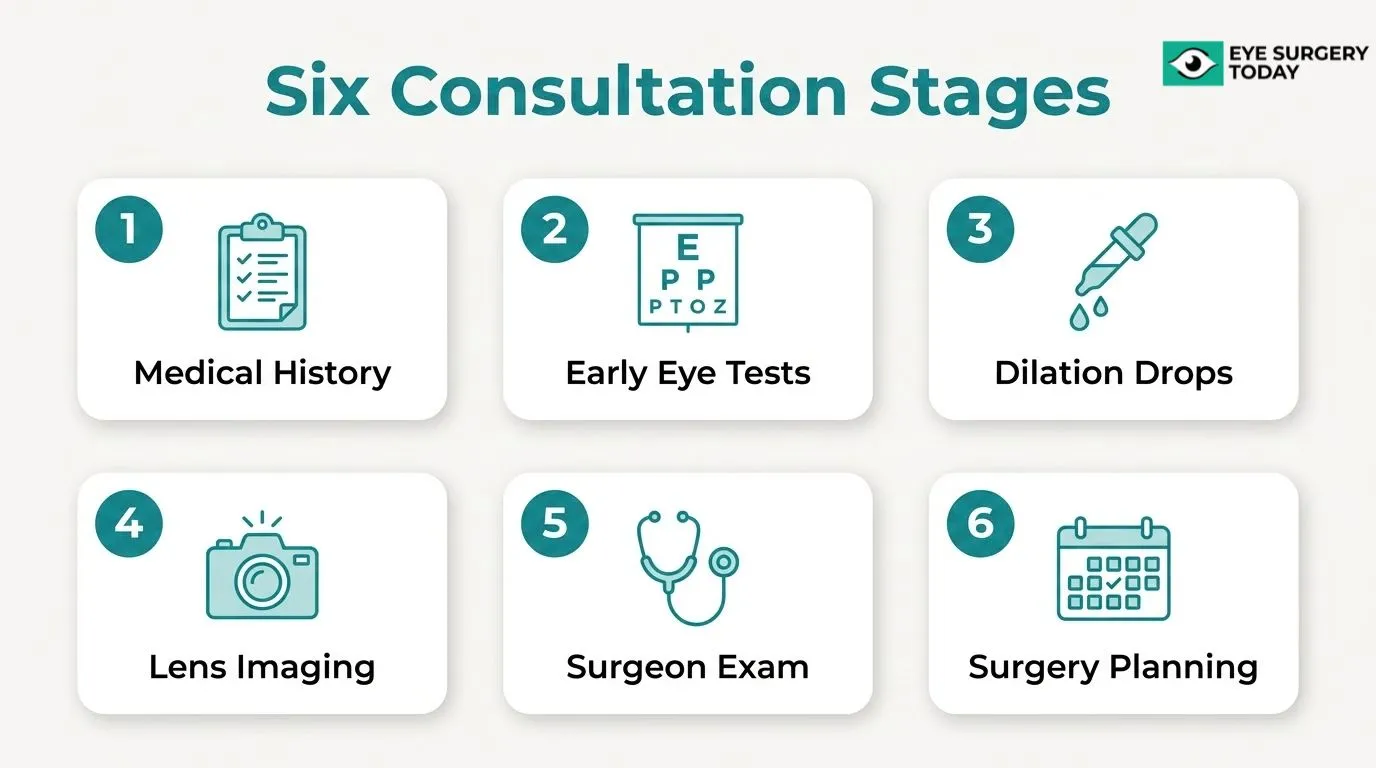
The consultation follows a structured sequence: intake and medical history review, early vision tests, dilating drop administration, lens imaging, the surgeon’s assessment, and a pre-surgery planning conversation. Each professional you see (healthcare assistant, ophthalmic nurse, and surgeon) handles a distinct phase of this process.
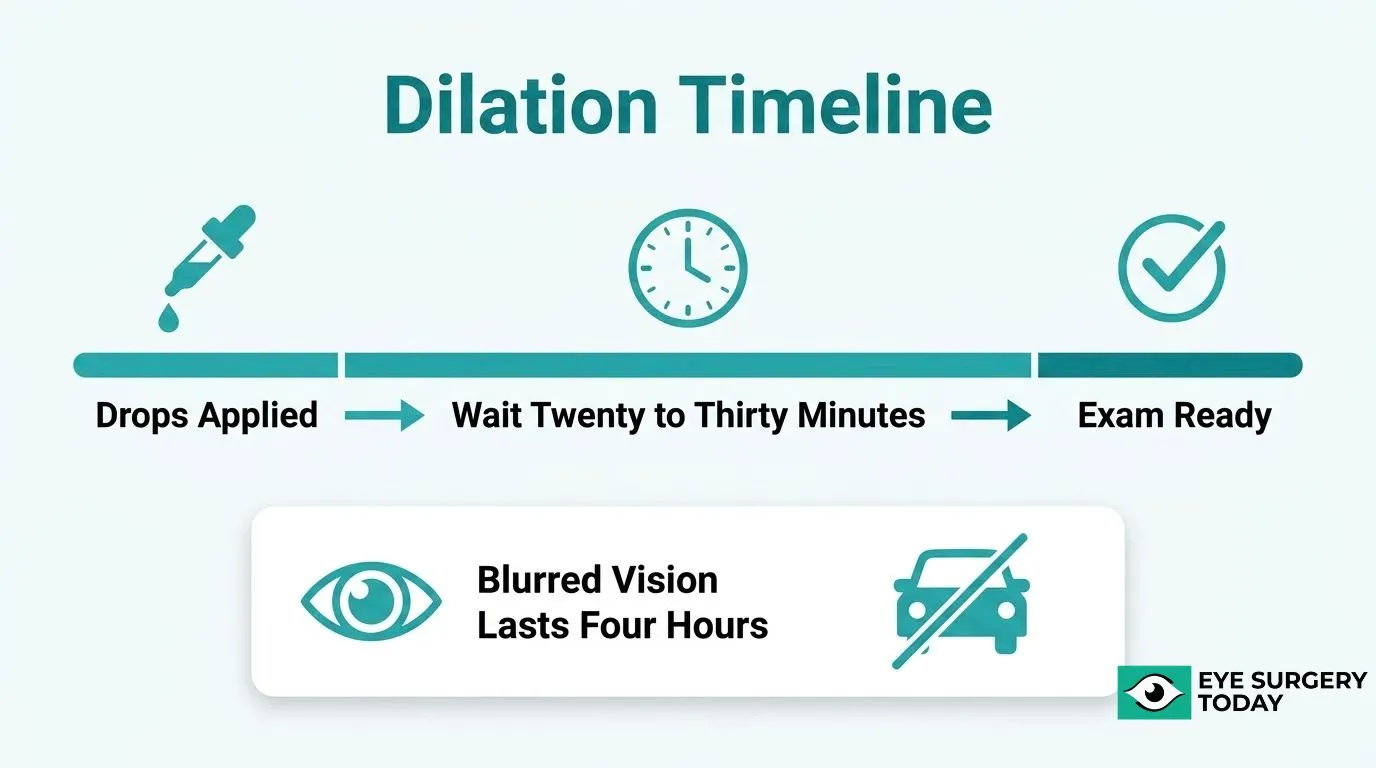
Dilation alone accounts for a large portion of the wait. Drops require roughly 20 to 30 minutes to reach full effect, and the blurred vision they cause can persist for several hours, which is why arranging a driver beforehand is essential.
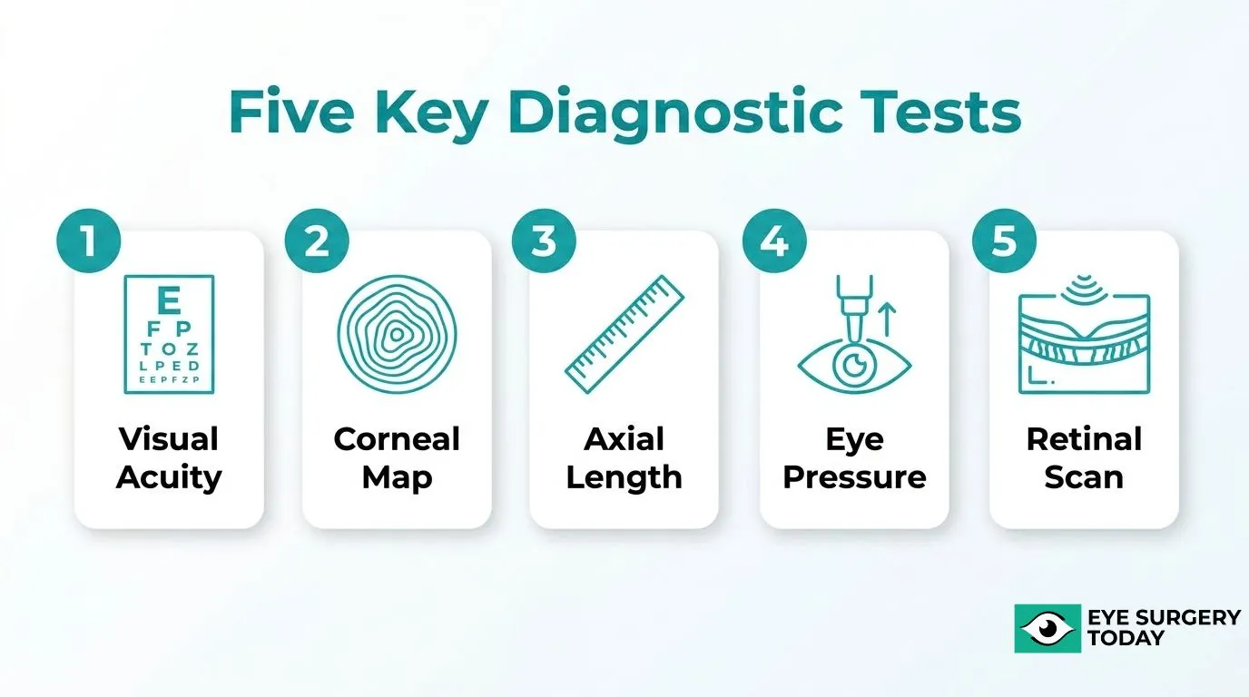
The diagnostic stage includes visual acuity testing, corneal topography, axial length measurement, intraocular pressure checks, and retinal imaging with OCT. These measurements feed directly into IOL power formulas that calculate the correct lens strength for your eye.
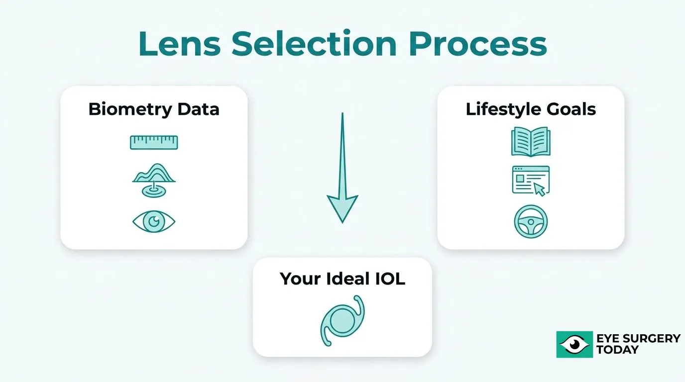
Lens selection combines biometric data with a lifestyle discussion about your visual priorities, covering monofocal, extended depth of focus, and other premium options. After the consultation, insurance authorization, a fitness-for-surgery review, and anesthesiology clearance must be completed before a surgery date is confirmed.
What Actually Happens During a Cataract Consultation?
A cataract consultation moves through several structured stages, each contributing to the total 2-to-3-hour appointment time. The sections below cover intake, early eye tests, dilation, lens imaging, the surgeon’s assessment, and pre-surgery planning.
What Does the Intake and Medical History Review Involve?
The intake and medical history review involves collecting your ocular history and completing a comprehensive eye exam before any testing begins. According to Centers for Medicare & Medicaid Services (CMS) guidance, for patients whose only diagnosis is cataracts, the standard pre-operative workup requires one comprehensive eye exam and a single IOL power scan, such as an A-scan. Staff will record current medications, prior eye surgeries, and any systemic health conditions that could affect surgical planning. This stage may appear brief, but accuracy here directly shapes which tests follow.
What Eye Tests Are Performed at the Start of the Visit?
The eye tests performed at the start of the visit typically include visual acuity testing, intraocular pressure measurement, and an initial slit-lamp examination of the anterior segment. According to Moorfields Eye Hospital NHS Foundation Trust, dilating eye drops are usually administered early in the visit because they take 20 to 30 minutes to take effect and can cause blurred vision for approximately four hours. Starting drops early allows the clinical team to run these initial assessments while waiting for full dilation, making efficient use of appointment time.
How Long Does the Dilated Eye Exam Take?
The dilated eye exam takes approximately 20 to 30 minutes for the drops to reach full effect, with the examination itself adding additional time on top. Dilation is essential because it widens the pupil enough for the clinician to examine the lens, retina, and optic nerve clearly. The blurring effect can persist for up to four hours after the drops are instilled, which is the primary reason patients are advised not to drive themselves home from the consultation.
What Happens During the Lens Measurement and Imaging Stage?
The lens measurement and imaging stage is where biometry data is collected to calculate the correct intraocular lens (IOL) power for your eye. According to the John A. Moran Eye Center at the University of Utah, Scheimpflug imaging generates axial, tangential, elevation, refractive, pachymetry, and difference maps alongside a 3D corneal model, while Placido disc-based topography provides accurate keratometry and astigmatism measurements. These scans are painless and non-contact. The Royal Free London NHS Foundation Trust notes that during this assessment stage, the cataract team discusses your vision goals, specifically whether you want better distance or near vision, since this preference directly influences which IOL is selected.
When Does the Surgeon Come In and What Do They Assess?
The surgeon comes in after the preliminary tests and imaging are complete, reviewing all biometry data and conducting a direct examination of the eye. Harvard Medical School’s Department of Ophthalmology recommends using high-quality optical biometry for greatest accuracy, though A-scan ultrasound biometry may be necessary for dense white or brunescent cataracts that prevent optical measurement. The surgeon also completes a perioperative risk assessment. According to NHS England, patients are not given a surgery date until they have been confirmed as medically fit for the procedure.This review stage is when the surgeon identifies any complicating factors that could affect surgical technique or lens selection.
What Is Discussed During the Pre-Surgery Planning Conversation?
The pre-surgery planning conversation covers the surgical approach, anesthesia type, lens options, recovery expectations, and follow-up scheduling. Common questions addressed at this stage include what to expect from anesthesia, whether any current medications need to be stopped, what post-operative activity restrictions apply, and when driving can resume. The surgeon will also confirm whether a follow-up visit is required and at what interval. This discussion is where patients should raise all remaining concerns before a surgery date is confirmed, making it one of the most decision-critical parts of the entire consultation.
Why Does Eye Dilation Add So Much Time to the Appointment?
Eye dilation adds time because the pupils require roughly 20 to 30 minutes to widen fully after drops are administered, and the dilated state must be maintained for the surgeon to examine the retina and lens clearly. The waiting period is unavoidable and typically runs concurrent with other parts of the consultation.
How Long Does Dilation Actually Take?
Dilation takes approximately 50 minutes from drop administration to the point when the pupil is ready for examination. According to a study published in the Journal of Ocular Pharmacology and Therapeutics via Washington University School of Medicine Digital Commons, the mean time between dilation and pupil examination was approximately 51 minutes regardless of the drop combination used. This waiting period is built into the appointment structure, but it explains why patients often feel the visit is slow in the middle even when everything is running on schedule.
What Side Effects Does Dilation Cause After the Visit?
The side effects of dilation include blurred vision, light sensitivity, and an inability to drive safely. According to the American Academy of Ophthalmology, patients should avoid driving and operating machinery until pupil dilation wears off. Effects can persist for two to six hours. Patients should arrange a driver for the appointment, as leaving independently is not safe while dilation remains active.
What Eye Tests and Measurements Make the Consultation So Thorough?
The tests that make the consultation so thorough include visual acuity testing, corneal topography, axial length measurement, intraocular pressure testing, and retinal assessment. Each measurement serves a distinct purpose in building a complete picture of the eye before any surgical planning begins.
What Does Visual Acuity Testing Measure?
Visual acuity testing measures the smallest letters a patient can read on a standardized Snellen chart placed 20 feet (6 meters) away, tested one eye at a time, according to MedlinePlus from the U.S. National Library of Medicine. This baseline establishes how much the cataract is affecting functional sight in each eye independently. The result directly informs the urgency of surgery and documents the starting point against which postoperative improvement is later measured.
What Does Corneal Topography Reveal Before Surgery?
Corneal topography reveals the shape, curvature, and surface regularity of the cornea before surgery. Two common methods are used:
- Placido disc-based topography: Maps the anterior corneal surface and provides accurate keratometry (K) and astigmatism measurements, though it does not evaluate the posterior cornea.
- Scheimpflug imaging: Generates axial, tangential, elevation, refractive, pachymetry, and difference maps, plus a 3D corneal model, capturing both anterior and posterior surfaces.
Identifying irregular astigmatism or corneal surface abnormalities at this stage is essential, as these findings directly influence whether a toric or premium intraocular lens is appropriate.
Why Is Axial Length Measurement Critical for Lens Selection?
Axial length measurement is critical for lens selection because it quantifies the distance from the anterior corneal surface to the retinal pigment epithelium in millimeters, the single most important variable in determining the correct intraocular lens power. According to StatPearls from the National Library of Medicine Bookshelf, this measurement can be obtained using either optical biometry or ultrasound biometry, with optical biometry preferred for its accuracy. Even a small error in axial length translates into a meaningful refractive miss after surgery, making this one of the most consequential measurements taken during the consultation.
What Does Intraocular Pressure Testing Show?
Intraocular pressure (IOP) testing shows whether fluid pressure inside the eye falls within a safe range, which may help identify conditions such as glaucoma before surgery proceeds. The gold standard method is Goldmann applanation tonometry (GAT), which uses direct corneal contact, according to a report in the Journal of Clinical Medicine. A commonly used alternative is the non-contact applanation tonometer, often called the “air puff” tonometer, which flattens the cornea using a brief air pulse rather than direct contact. Elevated IOP detected at this stage may alter surgical planning or require treatment before cataract surgery can safely proceed.
How Is Retinal Health Assessed During the Consultation?
Retinal health is assessed using optical coherence tomography (OCT), which provides high-resolution, cross-sectional images of the ocular tissues at the back of the eye using light waves, according to Cambridge University Hospitals NHS Foundation Trust. This scan allows the surgeon to detect conditions such as macular degeneration, diabetic retinopathy, or epiretinal membrane before surgery. Undetected retinal disease can limit the visual improvement achievable after cataract removal, so this assessment is essential for setting realistic expectations and selecting the most appropriate lens type for each patient.
How Is the Right Intraocular Lens Chosen During the Consultation?
The right intraocular lens is chosen by combining precise biometric measurements with a structured conversation about the patient’s lifestyle and visual goals. The subsections below cover how IOL power is calculated, what questions guide lens type selection, and how surgeons present monofocal versus premium options.
How Is the IOL Power Calculated From Measurements?
IOL power is calculated by feeding biometric data into mathematical formulas that predict where the lens will sit inside the eye after surgery. The Barrett Universal II formula uses obligatory inputs such as the A-constant, axial length (AL), keratometry (K), anterior chamber depth (ACD), and target refraction, with optional inputs including lens thickness (LT) and white-to-white (WTW) distance. According to a review published in Ophthalmology and Therapy, the Hill-RBF 3.0 formula extends this further by also incorporating central corneal thickness (CCT) and gender as data inputs. Each formula interprets the same measurements slightly differently, so surgeons often run multiple formulas and compare results before finalizing a power selection.
What Questions Help Determine the Best Lens Type for a Patient?
The questions that help determine the best lens type for a patient center on lifestyle, prior eye history, and refractive goals. The surgeon uses biometry measurements obtained from a quick, painless scan to confirm which lens power suits the eye’s optical dimensions, as described by Gloucestershire Hospitals NHS Foundation Trust. Beyond measurements, the conversation covers which focal distance aligns with daily activities such as reading, driving, or screen use, and whether the patient wishes to minimize glasses dependence after surgery. The University of Hospitals Plymouth NHS Trust notes that the eye doctor will discuss and agree with the patient on the desired level of focusing to aim for with the lens implant. Patients with prior eye surgeries should bring those records, as previous procedures can influence formula selection and lens candidacy.
How Does the Surgeon Discuss Monofocal Versus Premium Lens Options?
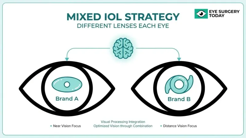
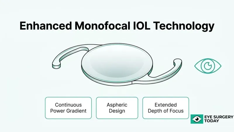
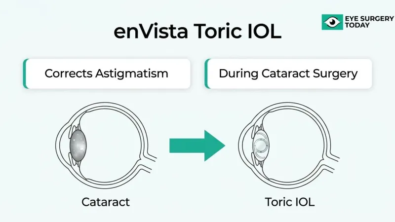
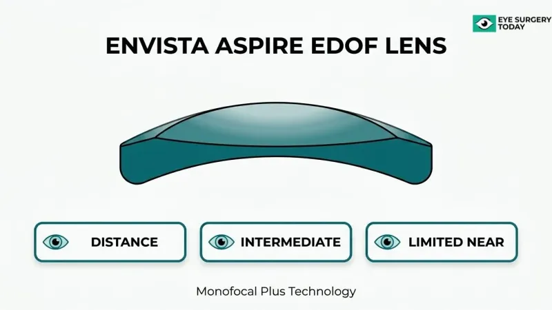
The surgeon discusses monofocal versus premium lens options by explaining the optical trade-offs of each category in relation to the patient’s daily visual demands. A monofocal lens corrects vision at one distance, typically either near or far, meaning glasses are usually still needed for the uncorrected range. According to the U.S. Food and Drug Administration, an Extended Depth of Focus (EDOF) intraocular lens uses optical design elements such as aspheric shaping, multiple refractive zones, or diffractive technology to provide a continuous range of relatively clear vision beyond what a monofocal lens achieves. Multifocal and toric IOLs represent additional premium categories, each addressing different patient needs such as presbyopia correction or corneal astigmatism. In practice, the lens discussion is one of the most consequential parts of the consultation; patients who arrive with a clear sense of their visual priorities tend to make faster, more confident decisions with their surgeon.
Who Else Will You Meet During a Cataract Consultation?
A cataract consultation involves more than just the surgeon. According to Torbay and South Devon NHS Foundation Trust, you will be seen by a healthcare assistant, an ophthalmic nurse, and an ophthalmic surgeon during the appointment. Each professional handles a distinct phase of your visit.
What Does the Healthcare Assistant Do?
The healthcare assistant typically manages intake tasks at the start of your visit, such as recording your basic health details, confirming your medication list, and administering dilating eye drops. This early groundwork allows clinical staff to focus on assessments rather than administrative tasks when you move through the appointment.
What Role Does the Ophthalmic Nurse Play?
The ophthalmic nurse performs several diagnostic tests, including visual acuity measurements and intraocular pressure checks, before the surgeon sees you. Nurses also explain each step of the process and monitor your comfort after dilation, which can cause temporary light sensitivity and blurred vision.
What Does the Ophthalmic Surgeon Assess?
The ophthalmic surgeon conducts the detailed clinical examination, reviews all test results, and leads the lens selection discussion. This is the stage where your suitability for surgery is confirmed and your refractive target is agreed upon. Meeting three professionals in sequence is the primary reason the appointment runs two to three hours.
What Should You Bring and Do to Prepare for the Appointment?
Preparing well for a cataract consultation saves time and helps the care team make accurate decisions. The key areas to organize in advance are transport, medical documentation, and a list of questions for your surgeon.
Why Should You Arrange a Driver Before the Appointment?
You should arrange a driver before the appointment because dilating eye drops will impair your vision for several hours after the consultation. According to Moorfields Eye Hospital NHS Foundation Trust, the drops can cause blurred vision for approximately four hours, making it unsafe to drive home. A study published in Ophthalmology (the American Academy of Ophthalmology journal) found that individuals with dilated pupils experienced increased glare-related issues and a higher incidence of collisions compared to those with undilated pupils. Arranging a driver in advance removes a logistical obstacle that could otherwise delay or disrupt the appointment.
What Medical Information Should You Bring With You?
The medical information you should bring includes your current medication list, insurance cards, a photo ID, and records of any previous eye surgeries. According to the University of Florida Department of Ophthalmology, helpful items include your medication list, a driver, insurance information, and prior surgical records if applicable. Bringing this information ensures the surgical team can verify drug interactions, confirm eligibility, and proceed with biometry and imaging without delays.
What Questions Should You Prepare to Ask the Surgeon?
The questions you should prepare to ask the surgeon cover lens selection, recovery, risks, and lifestyle fit. Having these ready before the appointment makes the surgical discussion more productive. Key questions to bring include:
- Which focal distance best matches my reading, computer use, and driving habits?
- How likely am I to still need glasses after surgery?
- Do I have any conditions, such as glaucoma, macular disease, or corneal astigmatism, that affect which IOL is suitable?
- What are the risks of cataract surgery?
- What type of anesthesia will I have, and will I feel pain?
- When will I be able to drive and resume normal activities after surgery?
- Will I need a follow-up visit with the surgeon after the procedure?
Preparing specific questions about your daily activities and visual priorities helps the surgeon recommend a lens type that genuinely matches your lifestyle.
What Happens After the Consultation Before Surgery Is Scheduled?
After the consultation, several administrative and clinical steps must be completed before a surgery date is confirmed. These include insurance authorization, additional pre-surgical testing, and an anesthesiology review.
According to Stony Brook Medicine’s Department of Ophthalmology, surgical coordinators obtain the necessary insurance authorization prior to surgery, and patients are directed to an Ambulatory Surgery Center (ASC) for further testing and a consultation with the anesthesiologist. Surgery scheduling depends on completing these steps and confirming medical fitness.
Patients should also expect a conversation about post-surgical vision outcomes, including whether glasses may still be needed for distance or reading after lens implantation. The answer depends on the IOL type chosen during the consultation: monofocal lenses correct one focal distance, so glasses are typically needed for tasks outside that range, while premium lens options may reduce that dependency for some patients.
From a practical standpoint, this post-consultation period is often where patients feel the most uncertainty. Understanding that the wait involves active clinical preparation, not just scheduling logistics, can make the process feel far less passive.
How Can Eye Surgery Today’s Guides Help You Prepare for Your Cataract Consultation?
A cataract consultation requires a specialist examination and eye measurements before any surgery date can be confirmed. Eye Surgery Today’s surgeon-reviewed guides and the key takeaways below help you arrive prepared and confident.
Can Eye Surgery Today’s Surgeon-Reviewed Resources Help Patients Understand Cataract Consultations?
Yes, Eye Surgery Today’s surgeon-reviewed resources can help patients understand cataract consultations by explaining each stage of the appointment before it happens. Cataract surgery is one of the most performed procedures globally: according to the 2023 National Ophthalmology Database Audit by the Royal College of Ophthalmologists, approximately 486,000 publicly funded cataract operations were undertaken in England and 13,000 in Wales during the 2021 NHS year alone.
Eye Surgery Today’s guides cover what to bring, such as a medication list, insurance cards, and photo ID, and what to expect from each assessment stage. Knowing the process in advance reduces uncertainty and helps patients ask better questions during their consultation.
What Are the Key Takeaways About Cataract Consultation Length We Covered?
The key takeaways about cataract consultation length are that the appointment is thorough by design, not by chance. NICE guidelines require that the referral decision involve a full discussion of how the cataract affects vision and quality of life, and that optical biometry be used to measure axial length for surgical planning.
The consultation covers medical history review, visual acuity testing, dilation, corneal imaging, biometry, intraocular pressure measurement, retinal assessment, lens selection, and pre-surgery planning. Each stage serves a specific clinical purpose and contributes to the 2 to 3 hour timeframe. Arriving prepared, with your medication list and a driver arranged, makes the process smoother and ensures the care team has everything needed to plan your surgery safely.