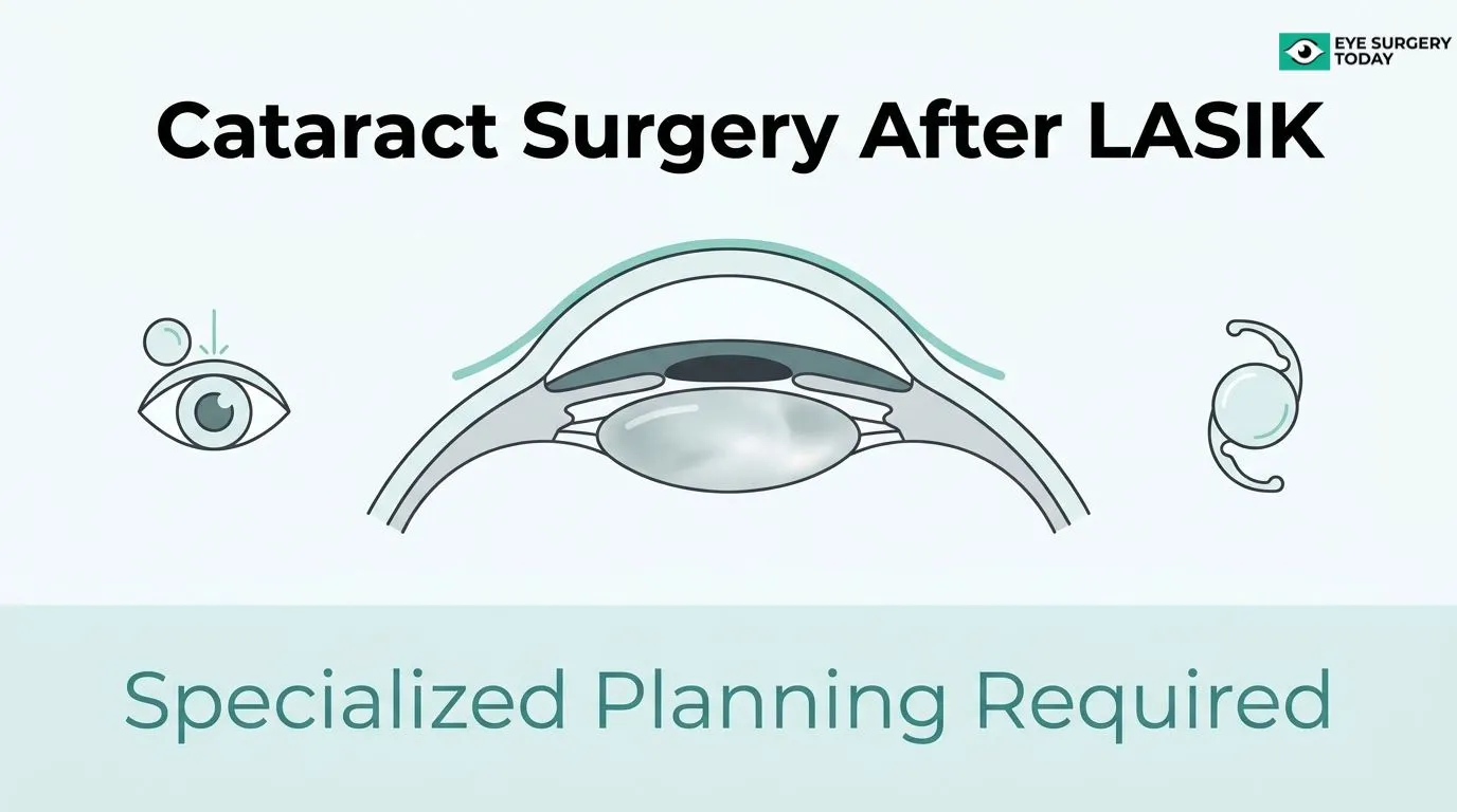
Cataract surgery after LASIK or PRK is a procedure that requires specialized IOL power calculation methods because prior corneal reshaping alters the measurements standard formulas depend on.
This guide covers how corneal changes from LASIK affect biometry accuracy, why historical surgical records play a critical role in planning, which IOL calculation methods and lens types surgeons consider for post-refractive eyes, the risks and visual trade-offs involved, and how patients can prepare for a more informed consultation.
LASIK permanently changes corneal curvature and disrupts the fixed anterior-to-posterior power ratio that conventional IOL formulas assume. This means standard keratometry may underestimate or overestimate true corneal power, leading to unexpected refractive error after cataract surgery.
Pre-LASIK keratometry, refraction records, and treatment maps give surgeons a baseline to calculate how much corneal power was removed. When these records are unavailable, no-history formulas and advanced tomography can partially compensate, though with wider prediction margins.
Specialized formulas like Barrett True-K and Haigis-L, along with intraoperative aberrometry, can improve IOL accuracy in post-LASIK eyes. Lens options range from monofocal to trifocal and EDOF designs; candidacy for premium lenses depends on corneal regularity, refractive stability, and retinal health.
Residual refractive error is more likely in post-LASIK eyes than in unoperated eyes, and visual phenomena such as halos or glare may be more pronounced with certain IOL designs. Staged enhancements and realistic outcome discussions before surgery can help manage these trade-offs.
Arriving at consultation with the right records, targeted questions, and calibrated expectations is one of the most effective ways to reduce refractive surprises in this inherently complex surgical scenario.
Why Does Prior LASIK Or PRK Make IOL Power Calculation More Complicated?
Prior LASIK or PRK makes IOL power calculation more complicated because these procedures permanently alter the cornea’s shape, refractive index, and curvature in ways that standard biometry formulas cannot reliably interpret. The following sections cover how myopic LASIK, hyperopic LASIK, PRK, and older surgical techniques each introduce distinct and clinically significant errors in lens power prediction.
How Does LASIK For Nearsightedness (Myopia) Affect IOL Calculations During Cataract Surgery?
LASIK for nearsightedness affects IOL calculations by flattening the central cornea, which causes standard keratometry instruments to underestimate true corneal power. This underestimation leads to selecting an IOL that is too weak, typically resulting in post-cataract hyperopia (farsightedness) the patient did not expect.
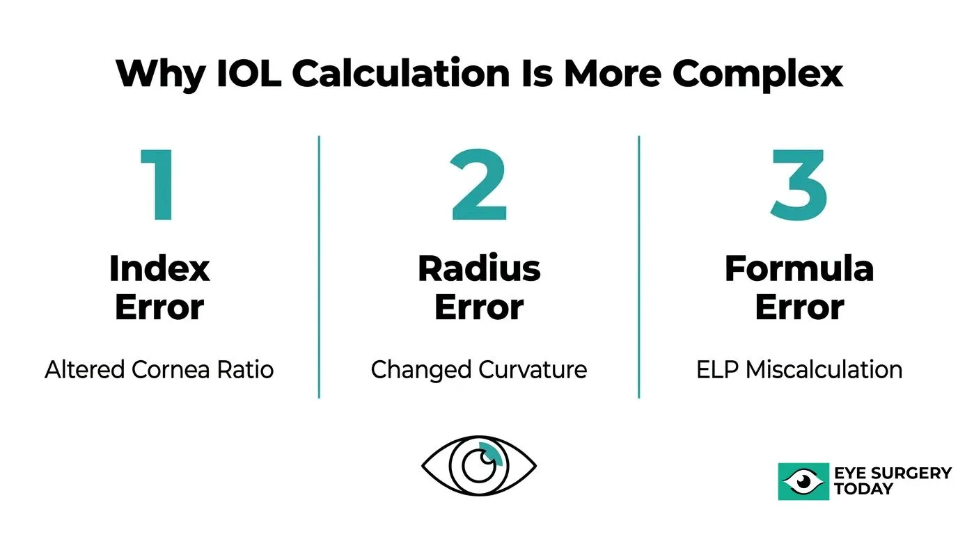
The core problem is threefold. According to the European Society of Cataract and Refractive Surgeons, the main sources of biometric error after corneal refractive surgery are miscalculation of the corneal refractive index, radius error, and formula error. Myopic ablation reduces the anterior corneal curvature while leaving the posterior cornea largely unchanged, breaking the fixed anterior-to-posterior ratio that standard formulas assume. Additionally, effective lens position (ELP) predictions, which rely on corneal curvature, become systematically inaccurate in flattened post-myopic eyes.
This combination of index error, radius error, and ELP error is why post-myopic LASIK eyes are among the most challenging scenarios in modern cataract planning.
How Does LASIK For Farsightedness (Hyperopia) Affect IOL Calculations During Cataract Surgery?
LASIK for farsightedness affects IOL calculations differently from myopic LASIK: it steepens the central cornea, causing standard keratometry to overestimate corneal power. This overestimation tends to push IOL selection toward a weaker lens than needed, often leaving patients with residual myopia after cataract surgery.
Hyperopic ablation also creates a distinct central corneal profile. Because the steepening is concentrated centrally, instruments measuring across a broader zone may capture a power reading that does not accurately reflect the optical axis. The result is a different direction of refractive surprise than myopic LASIK, but the underlying mechanism is the same: the altered anterior-to-posterior corneal power relationship invalidates the refractive index assumptions built into conventional IOL formulas.
Surgeons planning cataract surgery after hyperopic LASIK must account for this steepening bias and use post-refractive-specific formulas calibrated for this pattern.
How Does PRK Compare To LASIK In How It Complicates IOL Power Selection?
PRK complicates IOL power selection through the same three core mechanisms as LASIK: refractive index miscalculation, radius error, and formula error. The optical outcome of tissue removal is functionally equivalent, so both procedures produce the same categories of biometric challenge for cataract surgeons.
The meaningful clinical difference lies in corneal stability timing. The Asia-Pacific Journal of Ophthalmology notes that it is generally observed that the cornea takes longer to stabilize following PRK than LASIK. This prolonged stabilization period means that any pre-cataract measurements taken too soon after PRK may not reflect the cornea’s true final curvature, introducing an additional source of error that is less pronounced after LASIK.
In practice, if a patient has well-stabilized PRK with available pre-operative records, the calculation challenge is essentially parallel to LASIK. The instability variable is what sets PRK apart as a planning consideration.
How Do Older LASIK Techniques (Like Early Microkeratome LASIK) Impact Modern IOL Planning?
Older LASIK techniques impact modern IOL planning primarily through smaller and less predictable optical zones. Early microkeratome-based procedures often produced optical treatment zones smaller than 6 mm, and according to Cataract and Refractive Surgery Today Europe, when the optical zone diameter is smaller than 6 mm or the treatment is decentered, the corneal radius may be measured on the periphery of the treated zone rather than through the visual axis, introducing clinically significant radius error.
Beyond optical zone size, patients treated with early LASIK are also far less likely to have retrievable pre-operative keratometry or refraction records. Without historical data, surgeons must rely entirely on no-history formulas, which carry wider prediction error margins than history-based approaches.
Older techniques also more frequently produced corneal irregularities, which further limit the reliability of topographic measurements used in modern IOL planning workflows.
How Does Corneal Reshaping From LASIK Change The Eye’s Focusing Power?
LASIK reshapes the cornea to correct vision, but that reshaping permanently alters the measurements surgeons rely on to select an IOL for cataract surgery. The following sections explain how altered curvature disrupts biometry formulas, why standard keratometry loses reliability, and how shape changes translate into unexpected refractive error.
How Does Altered Corneal Curvature After LASIK Interfere With Standard Biometry Formulas?
Altered corneal curvature after LASIK interferes with standard biometry formulas primarily by disrupting the estimation of effective lens position (ELP). Most traditional IOL formulas predict ELP using corneal curvature as a proxy for anterior chamber depth. After LASIK flattens the cornea for myopia correction, that proxy becomes inaccurate, causing the formula to misplace the predicted IOL position and generate an incorrect power recommendation.
According to the Annals of Translational Medicine, the Haigis-L formula addresses this by deriving ELP from the actual anterior chamber depth rather than corneal curvature, using a correlation curve to compensate for both radius and keratometric index errors. This architectural change in the formula makes it considerably more robust for post-LASIK eyes than older generation vergence-based formulas that still rely on curvature-derived ELP estimates.
Why Do Standard Keratometry Readings Become Less Reliable After Corneal Refractive Surgery?
Standard keratometry readings become less reliable after corneal refractive surgery because the device samples only a small paracentral ring of the anterior corneal surface, missing the flattened central optical zone that LASIK creates. The instrument then extrapolates total corneal power using a fixed refractive index ratio between the anterior and posterior cornea, a ratio that no longer holds after stromal tissue has been selectively removed.
A study published in Clinical and Experimental Ophthalmology found that Pentacam 4.5-mm equivalent keratometry readings and simulated keratometry can serve as alternatives when pre-surgical data are unavailable, but clinicians must account for wide limits of agreement in subsequent calculations. In practice, this means even advanced topography-derived estimates carry meaningful uncertainty, and surgeons should treat any single keratometry value from a post-LASIK eye with deliberate caution rather than accepting it at face value.
How Does A Flatter Or Steeper Cornea After LASIK Lead To Unexpected Refractive Error After Cataract Surgery?
A flatter or steeper cornea after LASIK leads to unexpected refractive error after cataract surgery through three compounding mechanisms: refractive index miscalculation, radius measurement error, and formula selection error. As noted by the European Society of Cataract and Refractive Surgeons, these three sources are the primary drivers of biometric error following corneal refractive surgery.
The root of the refractive index problem is physical: as established in the Middle East African Journal of Ophthalmology, changing the anterior-to-posterior corneal power ratio alters the cornea’s effective refractive index in direct proportion to the amount of tissue removed. Radius error is a separate but related risk. According to Cataract and Refractive Surgery Today Europe, eyes treated with small or decentered ablation zones may have their corneal radius measured on the periphery of the treated zone rather than along the visual axis, introducing an additional mismatch. Optical zones of 6 mm or larger reduce this error to a negligible level.
To partially correct for formula error, the double-K method applies pre-surgical corneal power in the ELP prediction step while using the post-surgical reading for vergence calculation, as described by EyeWiki from the American Academy of Ophthalmology. Corneal stabilization timing adds further complexity: the Asia-Pacific Journal of Ophthalmology notes that the cornea generally takes longer to stabilize after PRK than after LASIK, meaning biometry performed too early in PRK patients may capture a still-changing corneal shape.
Understanding these layered error sources underscores why post-LASIK eyes require fundamentally different measurement and planning strategies before cataract surgery.
What Information From Your Old LASIK Or PRK Procedure Helps Plan Cataract Surgery?
Information from your old LASIK or PRK procedure helps plan cataract surgery by giving surgeons the pre-surgical corneal baseline needed to calculate IOL power accurately. The following H3s cover why pre-LASIK prescriptions matter, how original surgical records and treatment maps improve accuracy, and what surgeons can do when no historical data exist.
Why Are Pre-LASIK Glasses Or Contact Lens Prescriptions Helpful For IOL Selection?
Pre-LASIK glasses or contact lens prescriptions are helpful for IOL selection because they reveal the eye’s original refractive state before corneal reshaping altered its curvature. Standard biometry formulas estimate the cornea’s true power using a fixed refractive index, an assumption that breaks down after LASIK or PRK. Knowing the pre-surgical prescription allows surgeons to calculate how much corneal power was removed, giving a more reliable estimate of the eye’s original corneal curvature for use in IOL power formulas.
Without this baseline, surgeons must rely on approximations that introduce measurable error into the final lens selection. In practice, gathering this prescription is one of the simplest yet most impactful steps a patient can take before a cataract surgery consultation.
How Can Original LASIK Surgical Records And Treatment Maps Improve IOL Accuracy?
Original LASIK surgical records and treatment maps improve IOL accuracy by providing direct data on ablation depth, optical zone size, and pre- and post-operative keratometry. The American Academy of Ophthalmology Preferred Practice Pattern recommends that refractive surgeons maintain records of preoperative keratometry and refraction as well as postoperative refraction and make that data available for future care, including cataract surgery. Tangential curvature maps are particularly useful for determining the prior ablation pattern and reducing IOL calculation errors in eyes with a history of corneal refractive surgery, according to a case report published in the Journal of Refractive Surgery Case Reports.
Treatment maps also help identify small or decentered ablation zones, which can cause the keratometer to sample peripheral rather than central corneal tissue, introducing additional radius error.
What Can Be Done If No Historical LASIK Or PRK Data Are Available?
When no historical LASIK or PRK data are available, surgeons can use no-history IOL calculation formulas and advanced corneal imaging to estimate the eye’s pre-surgical baseline. A study published in the Journal of Cataract and Refractive Surgery found that in eyes with no historical data, the Barrett True-K no-history formula produced a significantly smaller median absolute refraction prediction error and a higher percentage of eyes within plus or minus 0.50 D compared to the Shammas and Haigis-L formulas. Pentacam-based equivalent keratometry readings offer an additional imaging-based alternative, though published studies note wide limits of agreement that must be factored into the calculation.
Combining multiple data sources, such as tomography, total keratometry, and intraoperative aberrometry, can partially compensate for the missing history, reducing but not eliminating residual uncertainty.
What Are The Main Methods Surgeons Use To Choose IOL Power After LASIK?
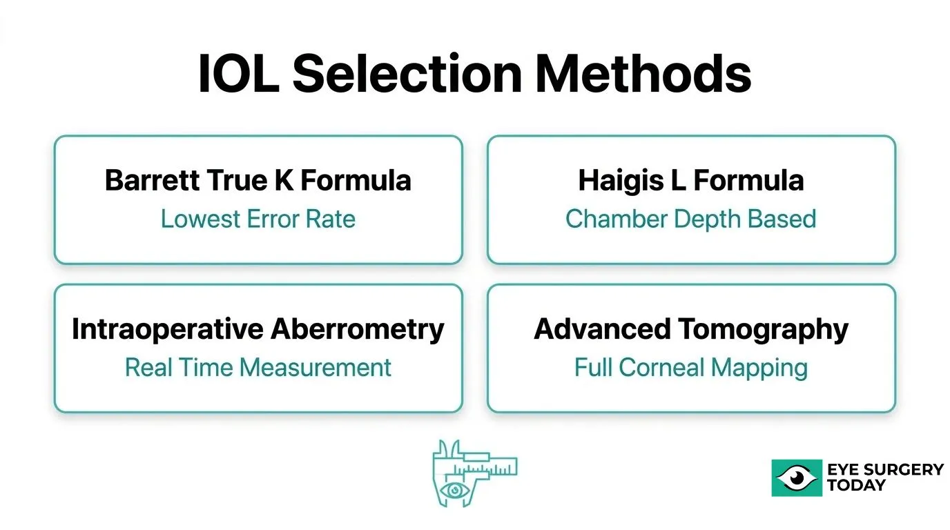
The main methods surgeons use to choose IOL power after LASIK include no-history calculation formulas, modern algorithm-based formulas, intraoperative aberrometry, and advanced corneal topography or tomography. Each approach addresses a different layer of the post-LASIK measurement challenge, and most surgeons combine two or more techniques to triangulate the safest IOL power.
How Do “No‑History” IOL Calculation Formulas Work For Post‑LASIK Eyes?
“No-history” IOL calculation formulas work by estimating corneal power without relying on the patient’s original pre-LASIK measurements. When historical records are unavailable, these formulas use regression-based algorithms or modified keratometric indices to reconstruct a reliable corneal power estimate.
A key adaptation used in many of these formulas is the double-K method. According to EyeWiki (American Academy of Ophthalmology), the double-K approach uses the post-refractive surgery corneal power reading for vergence calculation while substituting the pre-refractive surgery corneal power, or an estimate of it, for effective lens position (ELP) prediction. This separation prevents the altered cornea from compounding errors in two separate parts of the calculation.
When Are Modern Formulas Like Barrett True‑K Or Haigis‑L Preferred?
Modern formulas like Barrett True-K and Haigis-L are preferred when standard biometry formulas would produce clinically meaningful refractive surprises in post-LASIK eyes. Both are designed to correct the specific measurement errors that arise after corneal reshaping.
The Haigis-L formula compensates for radius and keratometric index errors through a correlation curve and derives ELP from anterior chamber depth rather than corneal curvature, reducing a major post-LASIK error source. According to a study published in Eye, Barrett True-K produced the lowest mean absolute error (0.424 D) among the formulas evaluated, outperforming Haigis-L (0.671 D) and Shammas-PL (0.638 D) across a post-LASIK cohort.
In eyes without historical data specifically, published findings in the Journal of Cataract and Refractive Surgery confirm Barrett True-K no-history achieved a significantly smaller median absolute prediction error than both Shammas and Haigis-L (both P < .05). For most post-LASIK cases, Barrett True-K is the stronger first-line choice.
How Can Intraoperative Aberrometry Help Fine‑Tune IOL Power During Cataract Surgery?
Intraoperative aberrometry helps fine-tune IOL power during cataract surgery by measuring the eye’s refractive state in real time, after the cataract has been removed but before the IOL is implanted. This live measurement bypasses the pre-operative corneal data errors that cause most post-LASIK miscalculations.
A study published in Clinical Ophthalmology evaluated 1,067 eyes and found that the ORA System achieved significantly higher proportions of eyes with absolute prediction errors of 0.25 D or less (47.2% vs. 42.5%, P = 0.0128) and 0.50 D or less (76.2% vs. 69.3%, P < 0.0001) compared with the Barrett True-K formula alone. Adding real-time aberrometry as a confirmatory layer on top of formula-based planning is particularly valuable for post-LASIK eyes, where pre-operative data uncertainty is highest.
What Role Do Advanced Corneal Topography And Tomography Play In Post‑LASIK IOL Planning?
Advanced corneal topography and tomography play the role of providing more complete corneal power data than standard keratometry, capturing posterior corneal curvature and surface irregularities that standard instruments miss. In post-LASIK eyes, where the anterior-posterior corneal relationship has been surgically altered, this additional data is clinically significant.
Pentacam-based measurements such as the 4.5-mm equivalent keratometry reading (EKR) and simulated keratometry may serve as alternatives to the clinical history method when pre-LASIK data are unavailable, though a 2010 study in Clinical and Experimental Ophthalmology notes that wide limits of agreement should be factored into any calculation. Total keratometry values, which incorporate posterior corneal measurements, feed directly into newer formula variants and can meaningfully reduce prediction error.
Using topography and tomography as the foundation for IOL planning, combined with formula selection and intraoperative aberrometry confirmation, represents the most defensible approach currently available for post-LASIK cataract cases.
What Types Of IOLs Are Commonly Considered After LASIK, And How Do They Differ?
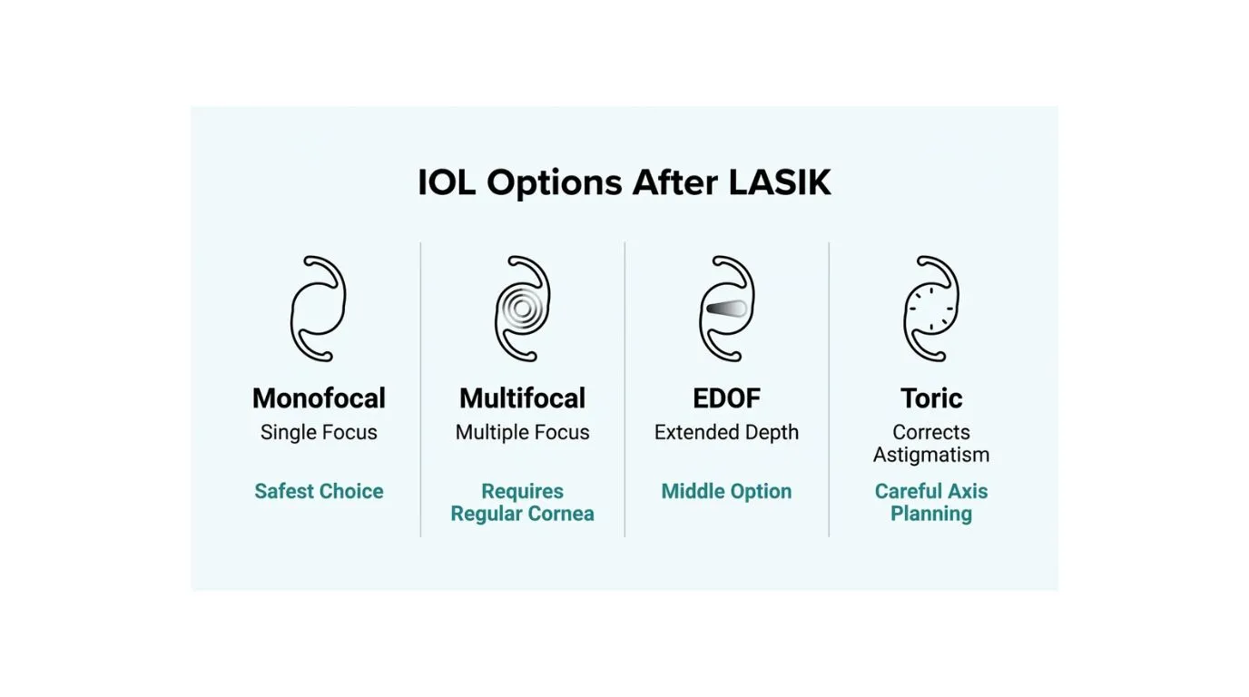
The types of IOLs commonly considered after LASIK include monofocal, multifocal, extended-depth-of-focus (EDOF), and toric lenses, each with different suitability profiles depending on corneal quality, residual astigmatism, and prior treatment characteristics.
When Might A Standard Monofocal IOL Be The Safest Choice After LASIK?
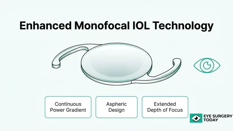
A standard monofocal IOL may be the safest choice after LASIK when corneal shape or power measurements are unreliable enough to make premium lens performance unpredictable. Monofocal lenses target a single focal point, typically distance vision, and their straightforward optical design tolerates residual refractive error better than multifocal designs. IOLs with negative spherical aberration can help offset the increased positive corneal spherical aberration that myopic LASIK commonly induces, according to a study published in Ophthalmology. For patients with irregular corneal profiles or insufficient pre-LASIK data, a monofocal lens with a planned mild myopic target is often the most reliable strategy to minimize unexpected outcomes.
Are Multifocal Or Extended‑Depth‑Of‑Focus IOLs Appropriate In Post‑LASIK Eyes?
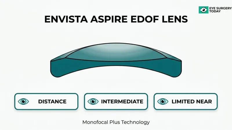
Multifocal and EDOF IOLs may be appropriate in carefully selected post-LASIK eyes, but only when corneal regularity and power calculations are sufficiently reliable. Trifocal IOL implantation can provide acceptable refractive and visual outcomes with minimal residual astigmatism in post-myopic LASIK and PRK eyes, according to research published in Clinical Ophthalmology. EDOF lenses may be particularly useful for patients who are not ideal multifocal candidates, as a study in the Journal of Ophthalmology found they can deliver efficient visual rehabilitation with good patient satisfaction and spectacle independence. In practice, EDOF designs are often a more forgiving intermediate option when some corneal irregularity remains but premium vision is still the goal.
How Does Prior LASIK Affect Candidacy For Toric IOLs To Correct Astigmatism?
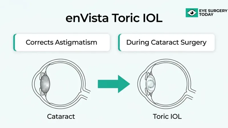
Prior LASIK affects toric IOL candidacy by altering corneal astigmatism patterns in ways that standard measurements may not fully capture. Toric lenses remain an excellent option for patients with residual astigmatism, but achieving optimal outcomes requires accounting for the posterior cornea’s against-the-rule astigmatism contribution and high angle alpha values, according to a 2024 review in Current Opinion in Ophthalmology. LASIK can shift the relationship between anterior and posterior corneal astigmatism, making standard axis-alignment assumptions less reliable. Surgeons typically rely on corneal tomography rather than simulated keratometry alone to plan toric IOL alignment in these eyes.
How Does Corneal Irregularity From LASIK Complications Limit Premium IOL Options?
Corneal irregularity from LASIK complications limits premium IOL options by degrading the optical quality needed for multifocal and EDOF lenses to function effectively. Irregular corneas scatter light unpredictably, amplifying the halos and dysphotopsias that premium lenses can already produce. Small or decentered LASIK treatment zones are a particular concern: research published in Cataract & Refractive Surgery Today Europe notes that when the optical zone diameter is smaller than 6 mm, corneal radius measurements may sample the peripheral treated zone rather than the visual axis, compounding IOL power errors. When significant irregularity is present, most surgeons consider a monofocal lens the only responsible choice.
Who May Be A Good Candidate For Premium IOLs After LASIK?
Good candidacy for premium IOLs after LASIK depends on corneal quality, refractive stability, retinal health, and personal visual goals. The following sections cover how each factor influences whether multifocal, EDOF, or toric lenses are appropriate.
How Does Overall Corneal Quality Influence Eligibility For Multifocal Or EDOF Lenses?
Overall corneal quality influences eligibility for multifocal or EDOF lenses by determining how cleanly light can be focused across multiple focal points. Corneas with well-centered LASIK ablation zones, minimal higher-order aberrations, and no significant irregularity are the strongest candidates. Irregular corneal surfaces scatter incoming light, which amplifies the halos and contrast-loss already associated with diffractive multifocal designs.
According to a study published in the Journal of Ophthalmology, EDOF IOLs may be a viable option for efficient visual rehabilitation with good patient satisfaction in eyes not primarily suited for multifocal implantation. This suggests EDOF lenses can serve as a practical middle ground when corneal quality falls slightly short of the strict threshold multifocals require. In practice, surgeons should evaluate corneal topography carefully before committing to any diffractive premium design.
Why Do Stable Refractions And Healthy Retinas Matter More In Post‑LASIK Cataract Planning?
Stable refractions and healthy retinas matter more in post-LASIK cataract planning because any residual refractive drift or macular pathology compounds the already elevated prediction error inherent to post-LASIK eyes. A refraction that has been unchanged for at least one year signals that the cornea has settled, giving biometric measurements their best chance of accuracy. Retinal conditions such as epiretinal membrane or early macular degeneration can undermine the contrast sensitivity that premium IOLs depend on, regardless of how accurately the lens power is calculated.
Dry eye disease also warrants attention at this stage. More than 90% of respondents in an ASCRS Clinical Survey reported that mild-to-moderate dry eye disease impacts patient satisfaction in cataract and refractive surgery, making preoperative ocular surface optimization an essential prerequisite.
How Do Lifestyle And Visual Priorities Shape IOL Choices After LASIK?
Lifestyle and visual priorities shape IOL choices after LASIK by clarifying which trade-offs a patient can realistically accept. Patients who drive frequently at night or work in low-contrast environments may find that multifocal designs introduce unacceptable halos or glare, making a monofocal or EDOF lens a more compatible fit. Conversely, patients with strong motivation to minimize spectacle dependence for reading and intermediate tasks may accept those trade-offs willingly.
Those with astigmatism remaining after LASIK should discuss whether a toric element is appropriate, since residual cylinder degrades the performance of any premium lens. A candid conversation about visual demands, tolerance for optical phenomena, and realistic outcomes is, in many ways, the most important step in premium IOL selection for any post-LASIK patient.
What Are The Potential Risks, Trade‑Offs, And Visual Side Effects In Post‑LASIK Cataract Surgery?
The potential risks, trade-offs, and visual side effects in post-LASIK cataract surgery include a higher likelihood of residual refractive error, halos, glare, and night-vision disturbances. The following sections explain why each occurs and what options exist if outcomes fall short of expectations.
Why Is There A Higher Chance Of Residual Refractive Error After Cataract Surgery In Post‑LASIK Eyes?
There is a higher chance of residual refractive error after cataract surgery in post-LASIK eyes because prior corneal reshaping makes IOL power prediction inherently less precise. The modified corneal shape alters how standard formulas estimate focusing power, introducing a measurable gap between intended and actual outcomes.
A study published in BMJ Open Ophthalmology found that differences between intended and postoperative refractive error were significantly greater in post-laser vision correction eyes than in control eyes (p=0.012), regardless of the calculation method used. Even advanced tools reduce, rather than eliminate, this gap. Surgeons who understand this limitation upfront can set more realistic refractive targets and plan enhancement options accordingly.
How Can Halos, Glare, And Night‑Vision Problems Change After Cataract Surgery In LASIK Patients?
Halos, glare, and night-vision problems can change after cataract surgery in LASIK patients because the pre-existing corneal profile interacts with the newly implanted IOL’s optics. Prior LASIK may have introduced spherical aberration or altered the transition zones of the cornea, which can intensify light-scatter symptoms when combined with certain IOL designs.
According to a study published in Life, up to 67% of patients may experience positive dysphotopsia immediately after cataract surgery, though only 2.2% report persistent symptoms at one year. For post-LASIK patients, the underlying corneal irregularity may extend the initial adjustment period or make premium IOL-related dysphotopsia more noticeable.
What Are The Options If Vision Is Not As Expected After Post‑LASIK Cataract Surgery?
The options if vision is not as expected after post-LASIK cataract surgery include spectacle or contact lens correction, laser enhancement procedures such as LASIK or PRK over the implanted IOL, and IOL exchange in select cases. The appropriate path depends on the magnitude and type of residual error, corneal thickness, and overall ocular health.
In practice, a staged approach is often the most prudent strategy: surgeons may target a slight myopic outcome intentionally in post-LASIK eyes, then refine with a surface laser enhancement once vision stabilizes. Patients benefit most when this contingency plan is discussed before surgery, not after an unexpected result has already occurred.
How Does The Cataract Surgery Process Differ For Someone Who Has Had LASIK?
The cataract surgery process differs for someone who has had LASIK in several important ways, from pre-operative testing to lens selection and recovery expectations. The sections below cover the extra measurements required, how surgical planning changes, and what patients can expect during recovery.
What Extra Pre-Operative Measurements And Tests Are Usually Needed?
The extra pre-operative measurements usually needed include corneal topography, tomography, total keratometry, and intraoperative aberrometry, in addition to standard biometry. Because prior LASIK alters the corneal shape, standard keratometry alone cannot reliably estimate true corneal power. Surgeons typically gather multiple data points across different devices to cross-check IOL calculations and reduce prediction error. Any available pre-LASIK records, including original keratometry and manifest refraction, are also requested to support history-based formula inputs.
How Might Surgical Planning And Lens Targeting Differ From A “Virgin” Cornea Case?
Surgical planning for a post-LASIK eye differs from a virgin cornea case primarily because the surgeon must account for corneal irregularities that affect how IOL formulas estimate lens position and power. Toric IOL planning requires particular care; according to a 2024 review in Current Opinion in Ophthalmology, achieving optimal satisfaction with toric lenses requires accounting for the against-the-rule astigmatism contribution from the posterior cornea and high angle alphas. Surgeons may also target a slight myopic outcome as a safety buffer, since post-LASIK eyes carry a higher risk of hyperopic surprise.
What Should Patients Expect During Recovery And Vision Stabilization After Surgery?
Patients should expect that vision stabilization after cataract surgery in a post-LASIK eye may take longer and follow a less predictable course than in eyes without prior corneal surgery. Initial vision may fluctuate during the first several weeks as the eye heals and the cornea settles around the new IOL. A small residual refractive error is more likely compared to non-LASIK eyes, and a spectacle prescription or enhancement procedure may be needed after the refraction stabilizes, typically around the six-week mark or later.
Understanding the full scope of these procedural differences helps patients enter consultation with realistic, well-informed expectations.
How Should You Prepare For A Cataract Surgery Consultation If You Had LASIK Before?
Preparing for a cataract surgery consultation after LASIK requires specific documentation, targeted questions, and an honest conversation about realistic outcomes. The following sections cover which records to gather, what to ask about IOL options, and how to calibrate expectations around glasses independence.
What Prior Eye Records And Prescription History Should You Bring To Your Surgeon?
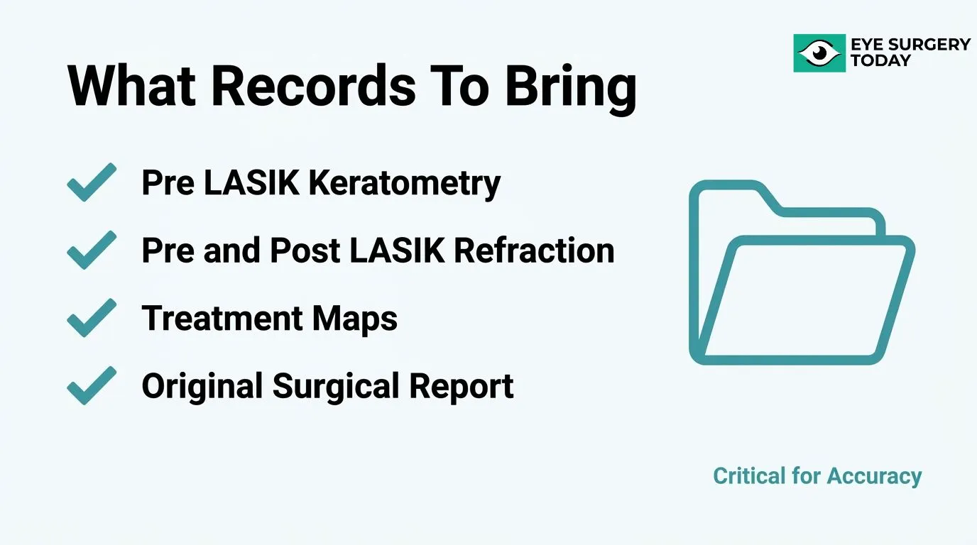
The prior eye records you should bring include your pre-LASIK keratometry measurements, your pre- and post-LASIK manifest refractions, and any corneal topography or treatment maps from your original procedure. According to the American Academy of Ophthalmology Preferred Practice Pattern, refractive surgeons are advised to maintain records of preoperative keratometry and refraction as well as postoperative refraction so that data are available for future care, including cataract surgery. Without this historical data, your surgeon must rely on estimation formulas, which introduce additional uncertainty into IOL power calculations. If your original LASIK clinic is still operating, contact them directly to request your full surgical file before your consultation.
What Specific Questions About IOL Options Should You Ask As A Post‑LASIK Patient?
The specific questions about IOL options you should ask as a post-LASIK patient center on formula selection, lens design, and contingency planning. Consider asking the following:
- Which IOL power formula will be used for your altered cornea, and why?
- Is intraoperative aberrometry available to refine lens selection during surgery?
- Are premium lenses such as trifocal or EDOF IOLs appropriate given your corneal topography, or is a monofocal the safer choice?
- If residual refractive error occurs, what enhancement options are available?
- Will your surgeon target slight myopia as a buffer against hyperopic surprise?
These questions help you understand the surgical plan and ensure your surgeon has accounted for the unique biometric challenges that prior LASIK creates.
How Can You Set Realistic Expectations About Glasses Independence After Surgery?
You can set realistic expectations about glasses independence by understanding that refractive predictability is lower in post-LASIK eyes than in eyes without prior corneal surgery. A review published by the National Library of Medicine confirms that accuracy and predictability remain inferior in eyes with a history of corneal refractive surgery, even with advanced formulas and imaging. This means a small residual prescription after cataract surgery is more likely than in first-time cataract patients, and full glasses independence is not guaranteed. Discussing a clear “Plan B,” such as a LASIK enhancement or lens exchange, before surgery gives you a practical framework if the primary result falls short of your goals.
How Do Cataract Surgery Outcomes In Post‑LASIK Eyes Compare With Eyes Without Prior Corneal Surgery?
Cataract surgery outcomes in post‑LASIK eyes compare less favorably than in eyes without prior corneal surgery, primarily in refractive predictability. The sub-sections below cover IOL prediction accuracy, patient satisfaction, and how emerging technology may narrow this gap.
What Do Studies Show About Accuracy Of IOL Predictions In Post‑LASIK Eyes?
Studies show that IOL prediction accuracy in post‑LASIK eyes remains measurably lower than in unoperated eyes. According to a review published on PubMed (National Library of Medicine), despite advancements in IOL power formulae, corneal imaging, and IOL options, accuracy and predictability remain inferior to eyes that undergo cataract surgery without a history of corneal refractive surgery.
Formula selection matters. A study published in the Journal of Cataract and Refractive Surgery found that, in eyes with no historical data, the Barrett True‑K no‑history formula produced a significantly smaller median absolute refraction prediction error and a greater percentage of eyes within ±0.50 D than both the Shammas and Haigis‑L formulas (P < .05).
Even with optimized formulas, achieving emmetropia in post‑LASIK eyes requires more planning than standard cataract cases.
How Do Satisfaction Rates And Spectacle Independence Compare Between The Two Groups?
Satisfaction rates and spectacle independence in post‑LASIK cataract patients can approach those of non‑LASIK patients when IOL type is matched carefully to corneal profile. A 2024 study published in BMC Ophthalmology found that 73% of post‑LASIK eyes and 75% of control eyes were within ±0.50 D of emmetropia at 6 months, with no statistically significant difference between groups (P > 0.05), and both groups achieved continuous satisfying visual acuity from near to distance.
For patients not well suited to multifocal IOLs, according to a study in the Journal of Ophthalmology, EDOF IOLs may provide efficient visual rehabilitation with good levels of patient satisfaction and spectacle independence.
Careful IOL selection, rather than corneal history alone, appears to be the stronger predictor of patient satisfaction in this population.
How Might Technological Advances Improve Outcomes For Future Post‑LASIK Cataract Patients?
Technological advances may improve outcomes for future post‑LASIK cataract patients through more precise corneal power measurement, smarter formulas, and better‑matched IOL designs. Tangential curvature mapping, for instance, can help determine the prior ablation profile and reduce IOL calculation errors in patients with a history of corneal refractive surgery, according to a case report in the Journal of Refractive Surgery Case Reports.
On the IOL design side, lenses with negative spherical aberration can reduce the positive corneal spherical aberration introduced by myopic LASIK or PRK, potentially improving optical quality. Research published in Clinical Ophthalmology also indicates that trifocal IOL implantation can provide acceptable refractive and visual outcomes with minimal residual astigmatism in post‑myopic LASIK and PRK eyes.
As these tools continue to mature, the outcome gap between post‑LASIK and virgin‑cornea cataract cases is likely to narrow further.
How Can Patient And Surgeon Work Together To Reduce Surprises After Cataract Surgery Post‑LASIK?
Reducing surprises after cataract surgery post-LASIK requires active collaboration between patient and surgeon across three areas: clarifying visual priorities before IOL selection, planning staged enhancements as a backup, and knowing when to seek specialist input.
Why Is A Detailed Discussion About Visual Priorities Essential Before Selecting An IOL?
A detailed discussion about visual priorities is essential before selecting an IOL because post-LASIK eyes carry a higher inherent risk of residual refractive error, making misaligned expectations a significant source of dissatisfaction. Patients should be specific about whether they prioritize distance clarity, near independence, or reduced halos and glare. These priorities directly influence whether a monofocal, EDOF, or trifocal lens is the most appropriate choice. Because predictive accuracy remains lower in post-LASIK eyes than in eyes without prior corneal surgery, surgeons need this information to plan a realistic refractive target from the outset. Patients who have not articulated their lifestyle needs are more likely to feel disappointed even when the surgical outcome is technically sound.
How Can Staged Or “Plan‑B” Enhancements Help Fine‑Tune Vision If Needed?
Staged or “Plan-B” enhancements can help fine-tune vision by correcting residual refractive error that persists after IOL implantation, using options such as glasses, contact lenses, LASIK enhancement, PRK touch-up, or lens exchange. Agreeing on an enhancement strategy before surgery reframes residual error as a manageable step rather than a failure. Surgeons may intentionally target a mild myopic endpoint in post-LASIK eyes to provide a buffer against hyperopic surprise, with enhancement available to refine the result. Discussing this pathway openly reduces anxiety and sets a realistic two-stage framework for achieving the best possible vision.
When Should A Second Opinion Or Referral To A Complex Cataract Specialist Be Considered?
A second opinion or referral to a complex cataract specialist should be considered when prior LASIK records are unavailable, when corneal topography shows significant irregularity, when the planned IOL is a premium lens in a high-correction post-LASIK eye, or when the patient’s visual goals are demanding. Surgeons who specialize in post-refractive cataract cases have access to advanced biometry tools, intraoperative aberrometry, and experience with edge-case formula selection that general cataract surgeons may use less frequently. Seeking a second opinion is not a sign of distrust; it is a practical step that can meaningfully narrow the gap between expected and actual visual outcomes.
How Should You Approach Cataract Surgery After LASIK With Independent Education From Eye Surgery Today?
Eye Surgery Today provides surgeon-reviewed education to help patients navigate complex decisions like cataract surgery after LASIK. The sections below cover how the platform’s resources support informed conversations with your surgeon and the key takeaways from this topic.
How Can Eye Surgery Today’s Cataract And IOL Education Help You Discuss Post-LASIK Options With Your Surgeon?
Eye Surgery Today’s cataract and IOL education can help you discuss post-LASIK options with your surgeon by building the foundational knowledge needed to ask the right questions, understand your measurement results, and evaluate lens recommendations confidently. Understanding why IOL calculations are more complex after LASIK, which formulas your surgeon may use, and what your pre-LASIK records contribute helps you arrive at a consultation prepared rather than reactive.
The American Academy of Ophthalmology Preferred Practice Pattern recommends that refractive surgeons maintain records of preoperative keratometry, refraction, and postoperative refraction, and provide that data for future care including cataract surgery. Knowing this standard exists means you can proactively request those records before your consultation, rather than discovering they are missing at a critical moment.
What Are The Key Takeaways About Cataract Surgery After LASIK And Why Prior Corneal Surgery Complicates Lens Selection We Covered?
The key takeaways about cataract surgery after LASIK center on one core truth: prior corneal reshaping introduces measurable layers of complexity into every stage of IOL planning, from biometry to lens selection to managing patient expectations.
The most important lessons from this topic include:
- LASIK alters corneal curvature in ways that make standard keratometry readings unreliable for IOL power calculation.
- Pre-LASIK surgical records, including keratometry and refraction data, meaningfully improve IOL accuracy when available.
- Specialized formulas such as Barrett True-K are better suited to post-LASIK eyes than standard formulas.
- Premium IOL options, including trifocal and EDOF lenses, may still be viable but require careful corneal quality assessment.
- Residual refractive error after cataract surgery is more likely in post-LASIK eyes, and realistic expectations about glasses independence are essential.
Eye Surgery Today’s surgeon-reviewed resources exist precisely for situations like this, where the gap between what patients know and what they need to know most affects their outcomes.