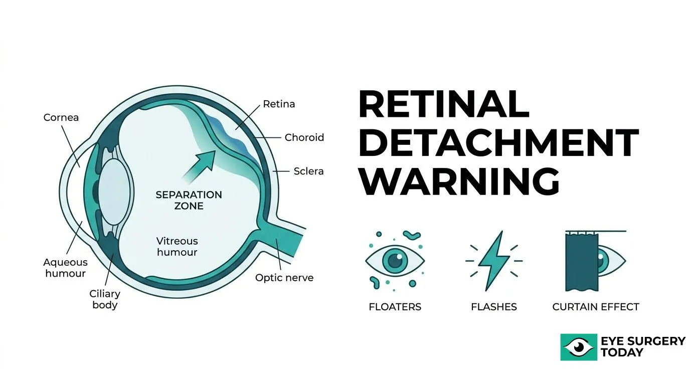
Retinal detachment is a serious eye condition in which the retina, the light-sensitive tissue lining the back of the eye, separates from its normal position. Without prompt treatment, this separation can lead to permanent vision loss.
This guide covers the warning signs and symptoms of retinal detachment, its causes and risk factors, the different types of detachment, how the condition is diagnosed and treated, and what patients can do to protect their vision through timely action and education.
The hallmark warning signs include sudden floaters, flashes of light, a dark curtain or shadow spreading across the visual field, blurry vision, and peripheral vision loss. Because retinal detachment is painless, these visual changes are often the only early indicators that something is wrong.
Several factors may contribute to retinal detachment, including age-related vitreous changes, eye trauma, high myopia, and prior cataract surgery. Risk levels vary depending on a patient’s individual profile; highly myopic individuals, for example, face substantially elevated rates compared to those without refractive error.
Three distinct types exist: rhegmatogenous (caused by a retinal tear), tractional (caused by scar tissue pulling the retina), and exudative (caused by fluid buildup without a tear). Each type follows a different mechanism and may call for a different treatment approach.
Diagnosis relies on a dilated fundus exam, sometimes supplemented by ultrasound or optical coherence tomography. Treatment options range from in-office laser or cryotherapy for retinal tears to surgical procedures such as pneumatic retinopexy, scleral buckling, and vitrectomy for more advanced cases. Recognizing symptoms early and understanding what to expect before and after treatment can help patients seek care within the critical window that may preserve long-term vision.
What Is Retinal Detachment?
Retinal detachment is a serious eye condition in which the retina, the light-sensitive layer of tissue at the back of the eye, pulls away from its normal position. According to the National Eye Institute (NIH), there are three types of retinal detachment: Rhegmatogenous, Tractional, and Exudative.
- Rhegmatogenous retinal detachment: caused by a retinal tear that allows fluid to seep beneath the retina.
- Tractional retinal detachment: results from scar tissue pulling the retina away, often linked to diabetic retinopathy.
- Exudative retinal detachment: occurs when fluid builds up under the retina without a tear, often due to inflammatory conditions such as uveitis.
Because retinal detachment can lead to permanent vision loss, recognizing its early signs is essential for protecting long-term eye health.
What Are the Warning Signs of Retinal Detachment?
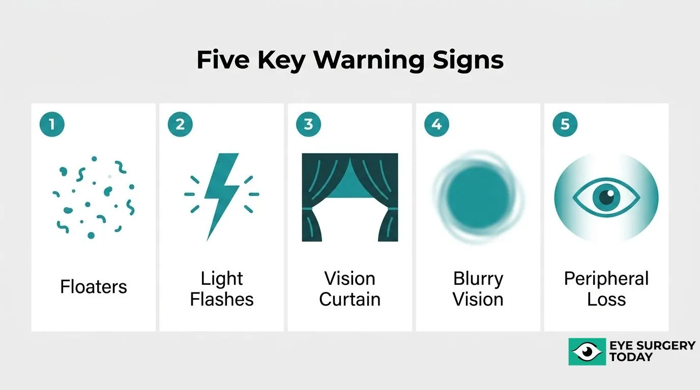
The warning signs of retinal detachment include sudden floaters, flashes of light, a dark curtain or shadow across vision, blurry vision, and peripheral vision loss. These signs can appear suddenly and require prompt evaluation by an eye care professional.
- Floaters
- Flashes of light
- Curtain or shadow in vision
- Sudden blurry vision
- Loss of peripheral vision
What Do Retinal Detachment Floaters Look Like?
Retinal detachment floaters typically look like small dots, lines, cobwebs, or specks drifting through the field of vision, often appearing suddenly and in greater numbers than usual. According to the NHS, these floaters commonly appear alongside flashes of light and a dark shadow in the vision. Retinal detachment itself causes no pain, so floaters may be the earliest noticeable warning sign a person experiences. A sudden surge of many new floaters differs from the occasional floater people notice with age, and this kind of change warrants prompt evaluation by an eye care professional rather than a wait-and-see approach.
What Do Flashes of Light From Retinal Detachment Feel Like?
Flashes of light from retinal detachment feel like brief, sudden bursts or streaks of light, often noticed in peripheral vision rather than straight ahead. These flashes tend to be short and small, sometimes described as lightning streaks or camera-flash sensations that appear and disappear quickly. According to Harvard Health Publishing, a new onset of floaters and flashes together is one of the key warning signs that vision trouble may be ahead. Flashes typically occur when the vitreous gel tugs on the retina, and they may grow more frequent as a retinal tear develops. Because flashes can precede a full detachment, noticing this symptom early gives patients a critical window for treatment before vision loss progresses.
What Does the Curtain or Shadow in Your Vision Mean?
A curtain or shadow in your vision means that part of the retina has likely detached and stopped sending visual signals from that section of the eye. Patients often describe it as a gray or dark veil descending from one edge of the visual field, gradually covering more area as detachment spreads. This symptom typically signals a more advanced stage of retinal detachment than floaters or flashes alone. According to UK HealthCare at the University of Kentucky, immediate evaluation by an ophthalmologist is recommended if a new floater appears, floaters accompany flashing lights, or part of the vision becomes dark. Because a curtain effect often means a larger area is already affected, it should be treated as an emergency rather than monitored at home.
Can Sudden Blurry Vision Be a Sign of Retinal Detachment?
Yes, sudden blurry vision can be a sign of retinal detachment, particularly when it appears alongside floaters, flashes, or a shadow in the field of vision. Blurring often develops as the detachment progresses toward the macula, the part of the retina responsible for sharp central vision. However, according to the American Society of Retina Specialists, some patients with retinal detachment may not notice any change in vision at all, which makes blurred vision an unreliable symptom on its own. Sudden changes in vision or eye pain that does not settle warrant urgent review, so blurry vision should never be dismissed as simple eye strain, especially when it appears suddenly rather than gradually.
Is Loss of Peripheral Vision a Warning Sign of Retinal Detachment?
Yes, loss of peripheral vision is a warning sign of retinal detachment and often indicates that the detachment has progressed beyond its earliest stage. Peripheral vision loss frequently appears as a shrinking or shadowed area at the edge of sight that gradually expands toward the center. According to Stanford University’s Mahajan Laboratories, peripheral vision loss, a sudden onset of new floaters, or a dark area moving across vision are all reasons to seek same-day evaluation from an eye surgeon. Left untreated, retinal detachment usually results in permanent severe vision loss or blindness, which is why peripheral changes should never be monitored passively at home. Recognizing this sign early, alongside floaters, flashes, and curtain-like shadows, gives patients the best chance of preserving vision through timely treatment.
With these signs identified, understanding what causes retinal detachment clarifies who faces the highest risk.
What Causes Retinal Detachment?
Retinal detachment develops from several distinct triggers that affect how the retina attaches to the back of the eye. Common causes include age-related vitreous changes, eye injury, high myopia, and complications following cataract surgery.
- Aging
- Eye injury
- Nearsightedness (high myopia)
- Cataract surgery
What Role Does Aging Play in Retinal Detachment?
Aging plays a significant role in retinal detachment because the vitreous gel inside the eye naturally shrinks and pulls away from the retina over time. This process, known as vitreous detachment, can create tiny retinal tears that allow fluid to seep behind the retina and cause it to separate. Because this shrinkage becomes more common with advancing age, older adults face a higher likelihood of experiencing this type of separation. This age-related process is considered a leading contributor to rhegmatogenous retinal detachment, the most common form of the condition, and it explains why routine eye exams become increasingly important later in life.
Can Eye Injury Cause Retinal Detachment?
Yes, eye injury can cause retinal detachment by physically tearing or separating the retina from the underlying tissue at the back of the eye. According to the Merck Manual Consumer Version, blunt injury may cause part of the retina, or the entire retina, to tear or separate from its underlying surface. This type of trauma often leads to rhegmatogenous retinal detachment, in which liquefied vitreous passes through a retinal break and lifts the retina away, appearing as undulating folds on examination. Sports injuries, falls, and blows to the eye or head are common examples of trauma associated with this type of detachment.
Does Being Nearsighted Increase Your Risk of Retinal Detachment?
Yes, being nearsighted can increase the risk of retinal detachment because elongated, myopic eyes place extra stretch on the retina and the surrounding vitreous gel. This added tension may hasten spontaneous posterior vitreous detachment (PVD), a common age-related condition whose prevalence tends to increase with age, according to research indexed on PubMed. When PVD occurs in an eye that is already elongated from myopia, the resulting traction on the retina can raise the likelihood of a tear or detachment. In clinical practice, this connection is one of the reasons eye care providers monitor highly myopic patients more closely for early retinal changes.
Can Cataract Surgery Increase the Risk of Retinal Detachment?
Yes, cataract surgery can increase the risk of retinal detachment, though the overall risk remains low for most patients. A large-scale analysis by Moorfields Eye Hospital NHS Foundation Trust found that among more than 3.1 million eyes studied, 6,690 (0.21%) developed rhegmatogenous retinal detachment within one year after cataract surgery. This modest increase in risk is generally linked to changes in the vitreous gel that can occur once the eye’s natural lens is removed and replaced with an artificial one. Patients with high myopia or prior eye trauma may face a somewhat higher risk following this procedure. Understanding these causes helps explain why detachment risk varies between patients.
What Are the Types of Retinal Detachment?
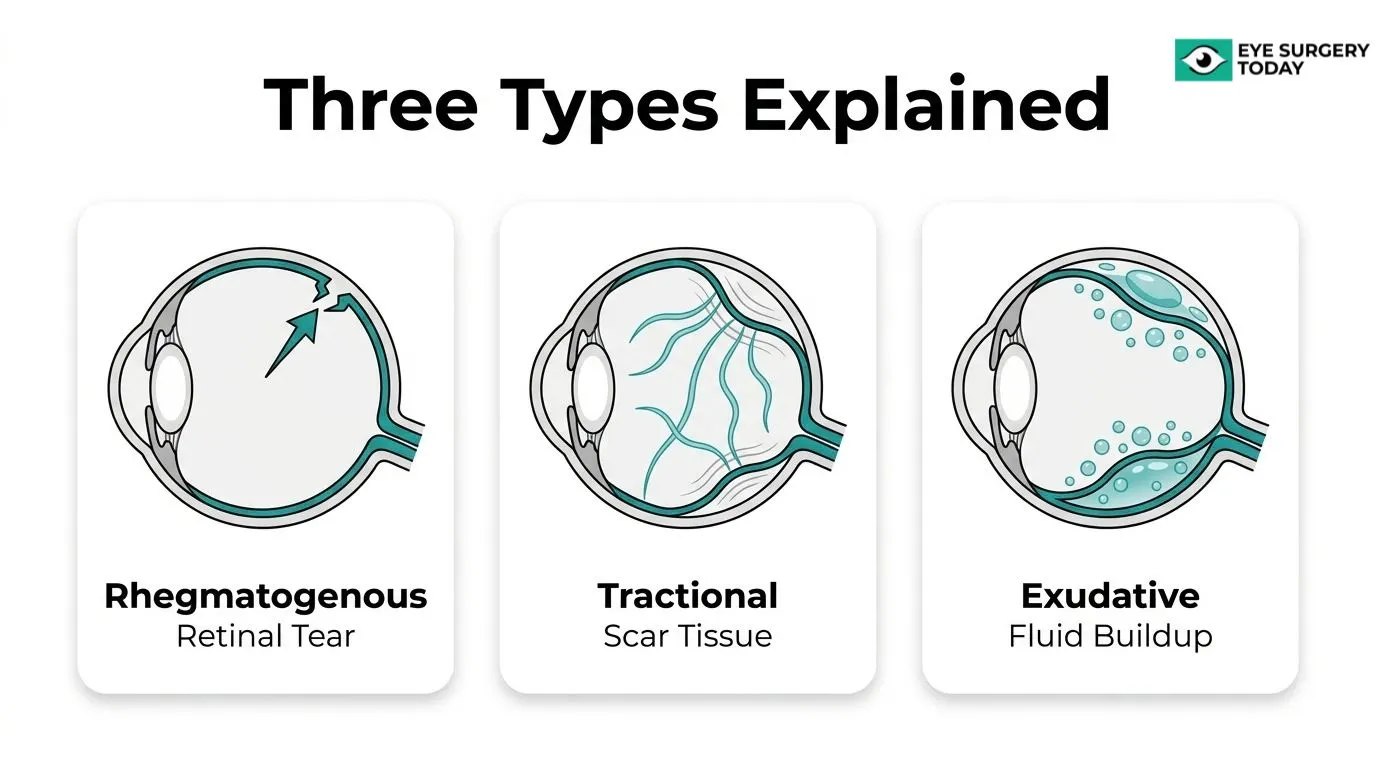
The types of retinal detachment are rhegmatogenous, tractional, and exudative, each defined by a different underlying mechanism that separates the retina from the tissue behind it.
- Rhegmatogenous retinal detachment
- Tractional retinal detachment
- Exudative retinal detachment
Rhegmatogenous Retinal Detachment
Rhegmatogenous retinal detachment is the most common form of retinal detachment, occurring when a retinal tear or break allows liquefied vitreous fluid to pass behind the retina and separate it from the retinal pigment epithelium. This differs from tractional retinal detachment, where mechanical traction pulls the retina away without any actual tear, a process often linked to neovascular conditions such as proliferative diabetic retinopathy, retinopathy of prematurity, or vitreomacular adhesions. According to a report from NewYork-Presbyterian on retinal detachment outcomes, rhegmatogenous retinal detachment occurs at a rate of 6.3 to 18 cases per 100,000 people each year. Because this type stems from a physical break in the retina, it often presents suddenly and typically calls for prompt evaluation by an eye care specialist.
Tractional Retinal Detachment
Tractional retinal detachment develops when scar tissue on the retina’s surface contracts and pulls the retina away from the back of the eye, without any retinal tear or hole present. This process usually stems from abnormal blood vessel growth, known as neovascularization, which can result from conditions such as proliferative diabetic retinopathy, retinopathy of prematurity, or vitreomacular traction. According to Columbia University’s Vagelos College of Physicians and Surgeons, tractional retinal detachment is most commonly associated with proliferative diabetic retinopathy. Because this type tends to progress gradually rather than suddenly, it can be easy for patients to underestimate its severity until vision loss becomes significant, which makes routine eye exams especially valuable for those living with diabetic eye disease.
Exudative Retinal Detachment
Exudative retinal detachment occurs when fluid, called exudate, builds up between the neurosensory retina and the retinal pigment epithelium without any retinal tear or traction involved. Unlike rhegmatogenous or tractional detachment, this fluid buildup stems from an underlying disease process rather than mechanical damage to the retina itself. According to the University of Iowa’s EyeRounds ophthalmology education program, inflammatory conditions such as uveitis are among the most common causes of exudative retinal detachment. Because no structural break occurs in this type, care typically focuses on addressing the underlying inflammatory or systemic condition rather than repairing the retina directly. Identifying which type is present helps guide who faces the greatest overall risk.
Who Is Most at Risk of Retinal Detachment?
The people most at risk of retinal detachment are those with high myopia, older adults, individuals with proliferative diabetic retinopathy, and residents of high-incidence regions.
High myopia is linked to substantially higher risk. A 2023 study in Scientific Reports found that incidence was 39-fold higher in highly myopic patients than in non-myopic patients (868.83 versus 22.44 per 100,000 person-years). Age and sex may also affect risk, as incidence peaks between ages 60 and 64, with men affected more than twice as often as women, a 2.3:1 ratio, according to research published in JAMA Ophthalmology. Proliferative diabetic retinopathy can raise risk as well, since abnormal blood vessel growth associated with the condition may pull the retina away from its normal position. Geography appears to play a role too, with global incidence averaging 12.17 per 100,000 people annually, ranging from 14.52 per 100,000 in Europe to 8.95 per 100,000 in the Americas.
Patients with overlapping risk factors, such as high myopia and diabetic retinopathy, often warrant closer monitoring by their eye care provider.
How Is Retinal Detachment Diagnosed?
Retinal detachment is diagnosed through a comprehensive, dilated eye exam performed by an eye care specialist. According to the American Academy of Ophthalmology, a dilated fundus exam is the critical first step, with B-scan ultrasound and optical coherence tomography (OCT) used as supplementary tools when the retinal view is limited. This exam typically includes:
- Pupil dilation: Eye drops widen the pupil for a full view of the retina.
- Fundus examination: A specialized lens lets the specialist inspect the retina for tears, folds, or fluid.
- B-scan ultrasound: This distinguishes retinal detachment from vitreous hemorrhage when the view is obstructed.
- Optical coherence tomography (OCT): This scan helps confirm subretinal fluid, especially near the macula.
How Is Retinal Detachment Treated?
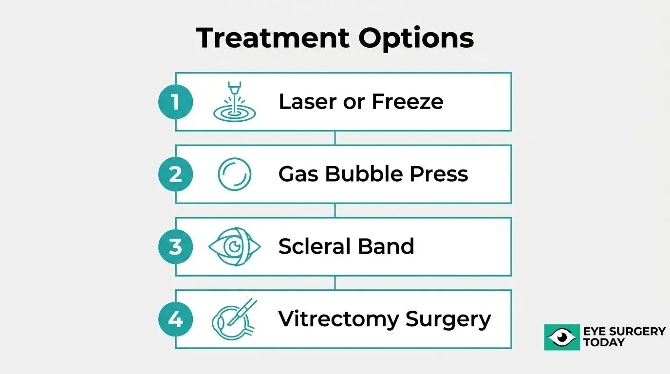
Retinal detachment treatment depends on the size, type, and severity of the detachment, ranging from simple office procedures to surgical repair. Common approaches include: 1) laser photocoagulation or cryotherapy for retinal tears, 2) pneumatic retinopexy, 3) scleral buckling surgery, and 4) vitrectomy.
What Happens During Laser or Cryotherapy for Retinal Tears?
During laser photocoagulation or cryotherapy, an ophthalmologist seals a retinal tear before it can progress to a full detachment. Laser treatment uses focused light to create small burns around the tear, forming a scar that binds the retina to the underlying tissue, while cryotherapy applies a freezing probe to the outer eye wall to achieve a similar effect. Both procedures are typically performed in an office setting under local anesthesia and tend to work best on tears with little or no fluid buildup underneath the retina. These treatments may help prevent a tear from advancing to a full detachment, though they are generally not suitable once the retina has already detached.
What Is a Pneumatic Retinopexy and When Is It Used?
Pneumatic retinopexy is a procedure in which an ophthalmologist injects a small gas bubble into the eye to press the detached retina back against the eye wall. Once the bubble holds the retina in position, laser or cryotherapy is used to seal the retinal break and secure the reattachment. This approach is generally reserved for certain rhegmatogenous detachments involving a single break located in the upper part of the retina. Patients may need to hold their head in a specific position for several days afterward to keep the bubble positioned correctly. Pneumatic retinopexy is less invasive than scleral buckling or vitrectomy, though it may carry a higher likelihood of needing additional treatment if the retina does not fully reattach.
What Does Scleral Buckling Surgery Involve?
Scleral buckling surgery involves placing a flexible silicone band around the outside of the eye to gently indent the eye wall and relieve the pulling force causing the retina to detach. The buckle remains permanently in place, supporting the area around the retinal break as healing occurs. According to a study published in the International Journal of Retina and Vitreous, primary reattachment rates for scleral buckling range from approximately 85% to 95%, with final success reaching 97% to 99% after reoperation. Surgeons often combine this technique with laser or cryotherapy to seal any breaks. This option is frequently considered for younger patients with clear vitreous and less complex detachment patterns, since it avoids some risks tied to intraocular surgery.
What Is Vitrectomy and How Does It Repair a Detached Retina?
Vitrectomy is a surgical procedure in which a surgeon removes the vitreous gel from the eye and replaces it with a gas bubble or silicone oil to flatten the retina against the eye wall. The surgeon uses small instruments inserted through tiny incisions to relieve traction on the retina, drain subretinal fluid, and seal retinal breaks with laser. Vitrectomy is often chosen for larger or more complex detachments, including those involving significant scar tissue or multiple tears. According to Johns Hopkins Medicine, some patients with detached retinas must undergo surgery the same day or within 24 hours to help protect vision. Recognizing detachment symptoms early helps determine which treatment option may still be possible.
What Happens If Retinal Detachment Is Left Untreated?
If retinal detachment is left untreated, the separated retina can lose its blood supply, causing the light-sensing cells to die and vision to fade permanently. What begins as a treatable, localized detachment often spreads to involve the macula, the area responsible for sharp central vision, and can progress to total retinal detachment and blindness in the affected eye.
Common consequences of delaying care include:
- Permanent loss of central or peripheral vision
- Complete detachment of the retina from the eye wall
- Increased risk of proliferative vitreoretinopathy, scar tissue that can lower the success rate of later surgery
- Blindness in the affected eye
In clinical practice, timely evaluation often makes the difference between preserved vision and permanent loss.
How Quickly Do You Need to Act When Symptoms Appear?
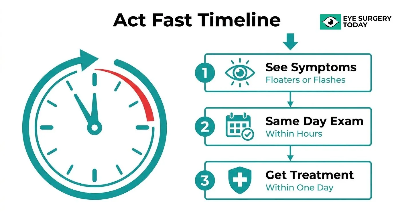
You need to act within hours, not days, once retinal detachment symptoms appear, because the retina’s blood supply is compromised the longer it stays detached. Timing depends on which warning sign shows up and how it progresses.
- New floaters or flashes with any vision field loss: According to the Royal Australian College of General Practitioners (Australian Family Physician), these symptoms require a same-day referral to an ophthalmologist for a dilated fundus examination to rule out a retinal tear or detachment.
- Confirmed retinal tear or detachment: Evaluation and treatment within 24 hours is generally considered the standard window before the detachment can spread further across the retina.
- Macula still attached (macula-on): This scenario is treated as the most urgent, since prompt surgery gives the central retina the best chance of staying protected.
- Macula already detached (macula-off): Research comparing outcomes by detachment duration suggests visual recovery may be more favorable when surgery happens within the first two to three days rather than later.
Retinal detachment is painless, so the visual symptoms themselves, not pain, are the only reliable trigger to seek same-day care. Waiting to “see if it gets better” is one of the most common and costly mistakes patients make with this condition.
Can Retinal Detachment Be Prevented?
Retinal detachment cannot always be prevented, but certain risk factors can be managed to lower the chances of it occurring. Prevention strategies vary by cause, such as protective eyewear for injury-related detachment, blood sugar control for diabetic retinopathy, and routine screening for people with high myopia or a family history of the condition.
- Eye injury: Wearing protective eyewear during sports, occupational tasks, or other high-risk activities may help guard against blunt trauma that can tear the retina, in line with guidance from the Occupational Safety and Health Administration on protecting eyes against mechanical hazards.
- High myopia or family history: Scheduling regular dilated eye exams allows an eye care provider to spot early retinal changes before a detachment develops.
- Age-related risk: According to the Centers for Disease Control and Prevention, adults over age 60 may benefit from a dilated eye exam every one to two years.
- Diabetic retinopathy: Managing blood sugar levels, as directed by a healthcare provider, may help reduce the abnormal blood vessel growth linked to tractional retinal detachment.
No strategy guarantees prevention, but recognizing new floaters, flashes, or a curtain-like shadow early and seeking prompt evaluation is generally considered the most reliable way to protect long-term vision.
How Does Eye Surgery Education Help Patients Recognize Retinal Emergencies?
Eye surgery education helps patients recognize retinal emergencies by turning clinical warning signs, such as floaters, flashes, and curtain-like shadows, into clear, actionable knowledge. The sections below explain how surgeon-reviewed resources support informed decisions and recap the key retinal detachment signs covered in this guide.
Can the Surgeon-Reviewed Guides at Eye Surgery Today Help You Understand Retinal Risks Before Surgery?
Yes, the surgeon-reviewed guides at Eye Surgery Today can help you understand retinal risks before surgery by clarifying which post-surgical changes are expected and which ones warrant concern. New floaters, for instance, are a normal finding after retinal surgery, particularly when gas has been used inside the eye to help reattach the retina, according to Moorfields Eye Hospital NHS Foundation Trust. Without this context, patients recovering from surgery may mistake a normal healing sign for a new emergency.
Eye Surgery Today’s surgeon-reviewed guides address this exact concern, helping patients tell the difference between expected postoperative floaters and symptoms that may signal a separate problem. Understanding this distinction before surgery is one of the most practical ways patients reduce anxiety during recovery, since knowing what to expect can prevent unnecessary worry over normal healing signs while still keeping patients alert to genuine red flags.
What Are the Key Takeaways About Signs of Retinal Detachment We Covered?
The key takeaways about signs of retinal detachment covered in this guide are that certain visual changes should never be ignored and that recognizing them quickly protects vision.
- Retinal detachment is typically painless, so vision changes, not pain, are the primary warning signal.
- New floaters, sudden flashes of light, and a dark curtain or shadow across the vision are the three classic warning signs.
- Peripheral vision loss and sudden blurry vision can also indicate a detaching retina, even without pain.
- Prompt evaluation by an eye care professional is essential whenever these symptoms appear suddenly, since delayed treatment raises the risk of permanent vision loss.
Recognizing these signs early, and understanding what is normal after any related eye surgery, gives patients the confidence to seek timely care when it matters most.