Vision worsening after eye surgery is a common patient concern that, in most cases, reflects temporary healing responses rather than a lasting complication. Corneal swelling, tear film instability, and postoperative medications can all reduce visual clarity during the early recovery window.
This guide covers the mechanisms behind temporary vision changes, normal recovery timelines by procedure, complications that may require intervention, warning signs that signal a medical emergency, and practical steps to support healing.
Several overlapping factors can make vision seem worse immediately after surgery, including corneal edema from endothelial cell disruption, dry eye caused by nerve and tear film changes, and the blurring effects of dilating drops like atropine. These responses are predictable and typically self-limiting as the eye stabilizes.
Recovery speed varies significantly by surgery type. Cataract surgery patients may notice functional improvement within days, though full stabilization can take weeks or longer. LASIK often restores usable vision within 24 to 48 hours, while vitrectomy patients may wait several months as an internal gas bubble gradually absorbs.
Though uncommon, complications such as posterior capsule opacification, cystoid macular edema, endophthalmitis, retinal detachment, and lens dislocation can worsen vision and may require additional treatment. Early detection through routine follow-up visits is associated with better outcomes for each of these conditions.
Recognizing red-flag symptoms is essential. Sudden vision loss, new flashes or floaters, escalating pain with blurriness, or worsening redness and light sensitivity all warrant same-day evaluation. Consistent use of prescribed eye drops and attendance at every scheduled follow-up appointment remain among the most effective ways to protect your visual recovery.
Why Might Vision Seem Worse Right After Eye Surgery?
Vision may seem worse right after eye surgery because of temporary healing responses, including corneal swelling, dry eye, and medication side effects. The following sections explain each cause.
Is Blurry Vision Normal in the First Few Days After Surgery?
Blurry vision is normal in the first few days after surgery. The eye needs time to heal from the surgical procedure, and several overlapping factors can temporarily reduce visual clarity during this window.
According to the American Academy of Ophthalmology, vision stabilization after cataract surgery typically occurs within 2 to 3 weeks, though it may take up to a year for the eye to feel completely normal. Most patients notice gradual daily improvement rather than an instant correction, so early blurriness rarely signals a problem.
Causes of short-term blurry vision after eye surgery include:
- Residual inflammation inside the eye from the surgical procedure.
- Mild corneal swelling that distorts how light reaches the retina.
- Pupil dilation from medicated drops used during and after surgery.
- Tear film instability on the ocular surface.
In most cases, this early blurriness resolves on its own as the eye heals. However, if vision worsens suddenly or is accompanied by severe pain, patients should contact their surgeon promptly rather than assuming it will pass.
How Does Corneal Swelling Affect Vision After Surgery?
Corneal swelling affects vision after surgery by distorting the way light passes through the cornea, the eye’s outermost refractive surface. When the cornea retains excess fluid, its normally transparent tissue becomes hazy, and incoming light scatters instead of focusing cleanly on the retina.
Post-operative corneal edema is primarily caused by endothelial pump failure resulting from mechanical trauma during phacoemulsification, chemical injury from irrigating solutions, or pre-existing endothelial compromise such as Fuchs endothelial corneal dystrophy, according to a review published through the National Center for Biotechnology Information.
Mild swelling typically resolves within the first week as endothelial cells restore their pumping function. Patients with pre-existing corneal conditions, however, may experience a slower recovery. Anti-inflammatory and hypertonic saline drops can help reduce fluid retention during this period. For most patients, this is one of the most predictable and self-limiting causes of early post-operative blur.
Can Dry Eye After Surgery Make Vision Seem Worse?
Yes, dry eye after surgery can make vision seem worse. An unstable tear film creates irregular light refraction across the corneal surface, producing fluctuating blur, glare, and visual discomfort that patients often mistake for a surgical complication.
According to a 2025 review in Frontiers in Medicine, LASIK-induced dry eye disease is driven by a reduction in corneal sensitivity due to transection of the sub-basal nerve plexus during flap creation, which impairs the blink reflex and the lacrimal functional unit’s feedback loop. While this mechanism is specific to LASIK, cataract and other intraocular procedures can also disrupt the tear film through exposure, inflammation, and preservative-containing postoperative drops.
Patients can manage post-surgical dry eye with:
- Preservative-free artificial tears used frequently throughout the day.
- Avoiding dry, windy, or air-conditioned environments when possible.
- Following the prescribed anti-inflammatory drop schedule to reduce ocular surface irritation.
Because dry eye symptoms often overlap with other causes of blurry vision, reporting them at follow-up appointments helps the surgeon distinguish between a healing tear film and a more significant issue.
How Do Dilating Drops and Medications Cloud Vision?
Dilating drops and medications cloud vision by temporarily paralyzing the pupil’s constriction muscles and the ciliary muscle responsible for focusing. With the pupil held wide open, excess light enters the eye, causing glare and reducing the ability to focus at near distances.
According to a 2023 study published through the National Center for Biotechnology Information, the use of postoperative atropine eye drops has been shown to cause a greater and longer-lasting reduction in visual quality compared to patients who do not receive them. Atropine’s prolonged cycloplegic effect can keep vision blurred for days after each application, which catches many patients off guard.
Other commonly prescribed postoperative medications, such as steroid and antibiotic drops, may also contribute to transient visual haze through mild corneal surface effects or temporary refractive changes. These medication-related symptoms resolve once the drops are tapered or discontinued according to the surgeon’s schedule. Patients should never stop prescribed drops early to “clear up” their vision, as doing so risks inflammation or infection.
With these temporary causes understood, recognizing the expected recovery timeline helps patients distinguish normal healing from signs that need medical attention.
What Does the Normal Vision Recovery Timeline Look Like?
The normal vision recovery timeline varies by procedure, with most improvements occurring within days to weeks. The subsections below cover what to expect at 24 to 48 hours, one week, and full stabilization.
What Should You Expect in the First 24 to 48 Hours?
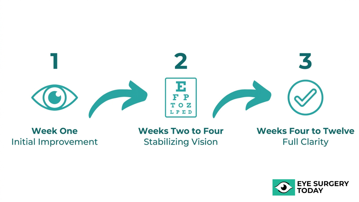
You should expect some degree of blurry, hazy, or fluctuating vision in the first 24 to 48 hours after eye surgery. This is the period when post-operative inflammation, residual dilation from surgical drops, and mild corneal swelling are at their peak. According to Dr. Agarwal’s Eye Hospital, functional vision recovery after LASIK typically occurs within 24 to 48 hours, whereas PRK requires several days for the epithelial layer to regenerate and several weeks for full visual clarity.
For procedures like cataract surgery this initial window often involves noticeably reduced sharpness. Colors may appear overly bright, and light sensitivity is common. These early symptoms, while understandably concerning, reflect the eye’s normal inflammatory response to surgical intervention rather than a complication. Rest, protective eyewear, and strict adherence to prescribed drops are the priorities during this phase.
How Clear Should Vision Be After One Week?
Vision after one week should be noticeably clearer than the first few days, though some residual blurriness or fluctuation is still considered normal. At this stage, much of the acute corneal swelling and inflammation has begun to resolve, and the eye is actively healing.
For LASIK patients, functional clarity is often well established by this point. Cataract surgery patients, however, may still experience mild haziness or shifting focus as the intraocular lens settles into position. PRK patients typically remain in the earlier phases of epithelial regeneration, so their vision at one week is often less sharp than LASIK or cataract patients at the same milestone. Patients who closely follow their post-operative drop schedule and avoid eye strain tend to notice more consistent improvement by this point.
When Does Vision Typically Stabilize Fully?
Vision typically stabilizes fully within a few weeks to several months, depending on the type of eye surgery performed. According to the American Academy of Ophthalmology, vision stabilization after cataract surgery typically occurs within 2 to 3 weeks, though it may take up to a year for the eye to feel completely normal. Following vitrectomy, vision typically begins to sharpen as the gas bubble shrinks between 4 to 6 weeks post-operatively, with continued improvement for up to three months.
Several factors influence the pace of full stabilization:
- Pre-existing conditions, such as diabetes or dry eye, can slow healing.
- The specific procedure performed determines the biological timeline for tissue recovery.
- Patient adherence to medications and follow-up visits supports optimal outcomes.
Any vision that worsens after a period of steady improvement warrants prompt evaluation, as this pattern can signal a complication rather than normal healing. Understanding these benchmarks helps set realistic expectations during recovery.
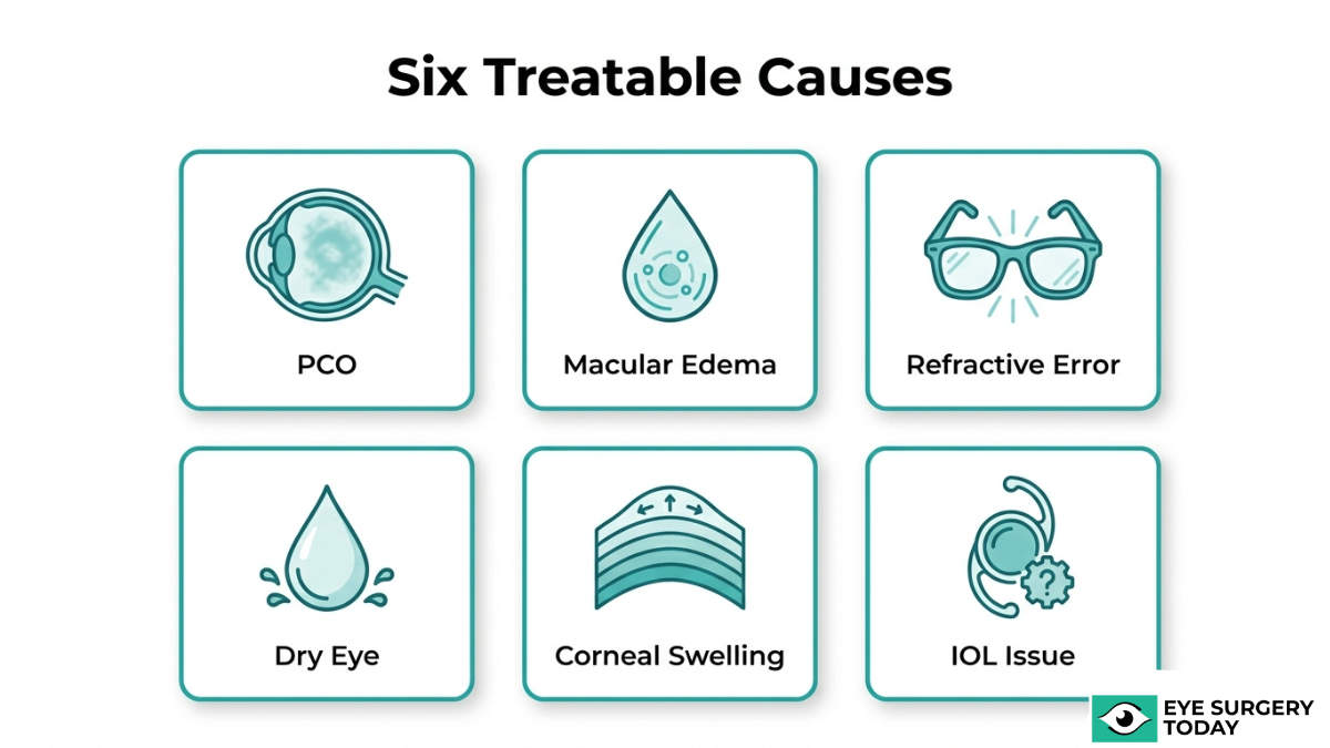
What Are Possible Complications That Can Worsen Vision?
Possible complications that can worsen vision after eye surgery include posterior capsule opacification, cystoid macular edema, endophthalmitis, retinal detachment, lens dislocation, and persistent corneal edema.
What Is Posterior Capsule Opacification?
Posterior capsule opacification (PCO) is a clouding of the thin membrane behind the intraocular lens that can develop weeks to months after cataract surgery. Often called a “secondary cataract,” PCO occurs when residual lens epithelial cells migrate and proliferate across the posterior capsule, scattering light before it reaches the retina. Symptoms typically include gradually increasing blur, glare, and reduced contrast sensitivity. A quick, painless Nd:YAG laser capsulotomy can treat PCO by creating a small opening in the opacified capsule. Because the onset is gradual rather than sudden, PCO is one of the more manageable complications; however, patients should not dismiss progressive haziness as normal and should report it promptly.
What Is Cystoid Macular Edema?
Cystoid macular edema (CME) is a condition in which cyst-like fluid-filled blisters accumulate within the macula, the central area of the retina responsible for sharp, detailed vision. Surgical inflammation can disrupt the blood-retinal barrier, allowing fluid to pool in cyst-like spaces that distort central sight. According to a large-scale registry study of U.S. patients published in the journal Ophthalmology (2016 to 2019), clinically significant CME occurred in 0.8% of eyes after cataract surgery. Patients with diabetes or a history of uveitis face elevated risk. Treatment typically involves topical anti-inflammatory drops, and most cases resolve within several weeks when identified early. Persistent CME, though uncommon, may require intravitreal injections to reduce swelling.
What Is Endophthalmitis?
Endophthalmitis is a severe intraocular infection that can develop after eye surgery when bacteria or, less commonly, fungi enter the eye. Symptoms often appear within days and may include rapidly worsening vision, increasing pain, significant redness, and eyelid swelling. Because the infection threatens the internal structures of the eye, endophthalmitis is considered an ophthalmic emergency requiring immediate treatment. Management typically involves intravitreal antibiotic injections and, in severe cases, a vitrectomy to remove infected vitreous fluid. Prompt recognition is critical; even a few hours of delay can significantly affect the visual outcome. Any sudden decline in vision accompanied by escalating pain in the early post-operative period warrants urgent evaluation.
What Is a Retinal Detachment After Eye Surgery?
A retinal detachment after eye surgery is a condition in which the retina separates from the underlying supportive tissue, interrupting its blood supply and threatening permanent vision loss. According to a study published by the National Center for Biotechnology Information, rhegmatogenous retinal detachment occurs in approximately 0.07% of eyes within one year following cataract surgery. Warning signs include a sudden increase in floaters, flashes of light, or the appearance of a shadow or curtain across part of the visual field. Patients with high myopia or a history of retinal tears face greater risk. Surgical repair, such as pneumatic retinopexy, scleral buckle, or vitrectomy, is typically required. Although rare, this complication demands same-day evaluation whenever symptoms arise.
What Is a Dislocated or Misaligned Intraocular Lens?
A dislocated or misaligned intraocular lens (IOL) is a shift of the implanted lens from its intended position inside the eye, causing blurred or double vision. According to research published by NCBI, IOL dislocation is most commonly late-onset, accounting for 89.16% of cases, with high myopia identified as a primary risk factor in 38.55% of affected patients. Weakening of the zonular fibers that support the lens capsule is the typical underlying cause. Symptoms may include fluctuating vision, visible lens edge, or monocular diplopia. Mild displacement can sometimes be monitored, while significant dislocation usually requires surgical repositioning or IOL exchange. Patients with pseudoexfoliation syndrome or prior ocular trauma should be especially attentive to gradual visual changes years after the original procedure.
What Is Persistent Corneal Edema?
Persistent corneal edema is prolonged swelling of the cornea that fails to resolve within the expected post-operative window, causing sustained blurry vision and discomfort. It is primarily caused by endothelial pump failure resulting from mechanical trauma during phacoemulsification, chemical injury from irrigating solutions, or pre-existing endothelial compromise such as Fuchs endothelial corneal dystrophy. When severe swelling prevents clear visualization through standard slit-lamp examination, anterior segment optical coherence tomography (ASOCT) serves as the preferred imaging tool for diagnosing complications like Descemet’s membrane detachment. Treatment ranges from hypertonic saline drops for mild cases to endothelial keratoplasty for eyes that do not recover adequate corneal clarity. Recognizing the underlying cause early gives surgeons the best chance of restoring comfortable, functional vision.
With complications identified, knowing when these symptoms become urgent guides timely action.
When Should You Worry About Vision Getting Worse?
You should worry about vision getting worse when specific warning signs appear after eye surgery. The following subsections cover sudden vision loss, new flashes or floaters, increasing pain, regression after improvement, and worsening redness with light sensitivity.
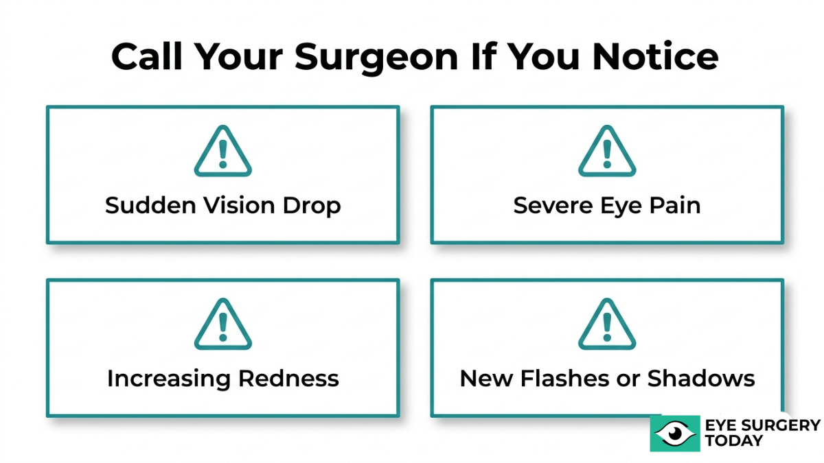
What If You Experience Sudden Vision Loss?
Sudden vision loss after eye surgery is a medical emergency that requires immediate evaluation. Unlike the gradual blurriness common during early recovery, a rapid or complete loss of sight in the operated eye may signal serious complications such as retinal detachment, endophthalmitis, or acute angle-closure glaucoma.
According to ophthalmologist Dr. Parth Shah, sudden vision loss, increasing pain, and redness are critical “red flag” warning signs of acute angle closure glaucoma, which is considered a medical emergency.
If vision drops significantly within minutes or hours, do not wait for your next scheduled follow-up. Contact your surgeon’s emergency line or go to the nearest emergency room. Early intervention in these scenarios can be the difference between preserving functional vision and permanent damage. Any sudden, unexplained decline in sight after surgery warrants same-day professional assessment.
What If You See New Flashes or Floaters?
New flashes or floaters after eye surgery can indicate traction on the retina, which may precede a retinal detachment. While a few floaters in the early postoperative period are not uncommon, a sudden shower of new floaters, flashing lights, or a curtain-like shadow across your visual field warrants urgent evaluation.
Rhegmatogenous retinal detachment occurs in approximately 0.07% of eyes within one year following cataract surgery, according to a study published on NCBI. Though the incidence is low, the consequences of a missed detachment are severe and often irreversible without prompt surgical repair.
A single new floater that remains stable may be benign. However, if multiple floaters appear suddenly alongside flashes of light, particularly in the peripheral vision, this pattern strongly suggests retinal involvement. Contacting your eye surgeon immediately gives you the best chance of preserving your vision.
What If Pain Increases Along with Blurry Vision?
Pain that increases along with blurry vision after eye surgery may indicate infection, elevated intraocular pressure, or severe inflammation. Mild soreness and a gritty sensation are typical during the first few days of recovery. Escalating pain, especially when accompanied by worsening vision, is not normal.
Endophthalmitis, a serious intraocular infection, can present with progressive pain and declining vision. According to the American Academy of Ophthalmology, acute postoperative endophthalmitis within 60 days of surgery has an incidence rate ranging from 0.058% to 0.964% depending on the surgical group and prophylaxis used.
The combination of increasing pain and visual deterioration is one of the most reliable clinical red flags in postoperative eye care. Rather than managing discomfort at home with over-the-counter medications, patients experiencing this pattern should contact their surgeon the same day. Delaying evaluation when pain and blurriness progress together significantly raises the risk of permanent vision compromise.
What If Vision Worsens After Initially Improving?
Vision that worsens after initially improving may signal a delayed complication rather than a normal fluctuation. When the operated eye shows clear progress during the first days or weeks, only to regress, conditions such as posterior capsule opacification, cystoid macular edema, or intraocular lens dislocation should be considered.
Posterior capsule opacification incidence ranges from 5% to 50% within the first year after cataract surgery, and Nd:YAG laser capsulotomy restores vision in over 95% of these cases immediately, according to a 2025 review published on ScienceDirect. This makes PCO one of the most treatable causes of secondary vision decline.
A regression pattern is particularly important to recognize because patients often assume the initial improvement means full healing has occurred. In clinical practice, any measurable decline in visual acuity after documented improvement should prompt a follow-up examination, even if the next scheduled visit is weeks away.
What If Redness and Sensitivity to Light Get Worse?
Redness and sensitivity to light that worsen after eye surgery may indicate uncontrolled inflammation, infection, or elevated intraocular pressure. Some degree of redness and photosensitivity is expected in the first postoperative week as the eye heals. When these symptoms intensify rather than gradually resolve, they become warning signs.
Persistent or worsening redness, particularly when combined with tearing, deep ocular ache, or hazy vision, can suggest uveitis, endophthalmitis, or a wound integrity issue. These conditions require prompt slit-lamp examination and possible treatment adjustment.
From a clinical perspective, worsening light sensitivity is an especially underappreciated red flag. Patients often attribute increasing photophobia to normal healing variability, but progressive sensitivity after the first week typically reflects active intraocular inflammation that may need more aggressive anti-inflammatory management. Report these changes to your eye care provider promptly rather than assuming they will self-resolve.
Understanding these warning signs prepares you to act quickly if complications arise after surgery.
What Should You Do If Vision Is Worse After Surgery?
If your vision is worse after surgery, you should contact your surgeon’s office promptly, attend a diagnostic evaluation, and discuss whether a corrective procedure may be needed. The following sections cover when to call, what tests to expect, and whether a second procedure can help.
Should You Contact Your Surgeon or Visit the ER?
You should contact your surgeon first for most post-operative vision concerns, as their office can assess whether symptoms fall within the normal healing timeline. Surgeons maintain after-hours call lines specifically for post-operative patients, making same-day or next-day triage possible in most cases.
However, certain situations warrant an emergency room visit instead:
- Sudden, complete loss of vision in the operated eye.
- Severe pain that worsens despite prescribed medications.
- Trauma or direct impact to the eye during recovery.
If your surgeon’s office is unreachable and symptoms are escalating rapidly, an ER with ophthalmology coverage can stabilize the situation until a specialist evaluation is arranged. When in doubt, calling your surgeon’s office first remains the safest starting point, since they have your surgical records and can guide the appropriate next step.
What Tests Might Your Doctor Perform?
Your doctor may perform several diagnostic tests to identify the cause of worsening vision after surgery. The specific tests depend on your symptoms and which procedure you had.
Common diagnostic evaluations include:
- Slit-lamp biomicroscopy to examine the cornea, lens implant position, and anterior chamber for signs of inflammation or structural issues.
- Optical coherence tomography (OCT) to capture cross-sectional retinal images and detect macular edema or other posterior segment changes.
- Anterior segment OCT (ASOCT) for cases where severe corneal swelling limits standard visualization.
- Intraocular pressure measurement to rule out post-surgical glaucoma.
- Dilated fundus examination to inspect the retina for detachment or other abnormalities.
According to an NCBI study on post-surgical corneal complications, ASOCT is the preferred imaging tool for diagnosing Descemet’s membrane detachment when severe corneal edema prevents clear visualization via slit-lamp biomicroscopy. These tests guide your surgeon toward the most effective treatment plan.
Can a Second Procedure Correct the Problem?
Yes, a second procedure can correct many post-operative vision problems, depending on the underlying cause. Not every complication requires additional surgery, but several common issues respond well to targeted interventions.
Corrective options include:
- Nd:YAG laser capsulotomy for posterior capsule opacification, a quick in-office procedure that restores clarity.
- IOL repositioning or exchange for a dislocated or misaligned intraocular lens.
- LASIK enhancement for residual refractive error after an initial refractive procedure.
- Vitrectomy for persistent floaters, macular conditions, or retinal detachment repair.
- Corneal procedures such as DSAEK or DMEK for cases of lasting corneal edema from endothelial cell damage.
The decision to pursue a second procedure typically comes after the eye has had adequate time to heal and stabilize. In clinical practice, most surgeons prefer to exhaust conservative management first, since many early vision issues resolve without further intervention. Your ophthalmologist can determine candidacy based on diagnostic findings and the specific nature of your complication.
Understanding your corrective options prepares you to discuss recovery expectations by surgery type.
How Does Vision Recovery Differ by Surgery Type?
Vision recovery differs by surgery type in timeline, post-operative care requirements, and the nature of temporary side effects. The sections below cover recovery specifics for cataract surgery, LASIK and PRK, and vitrectomy.
How Does Recovery Differ After Cataract Surgery?
Recovery after cataract surgery typically follows a gradual timeline, with most patients noticing functional improvement within days but full stabilization taking longer. According to the American Academy of Ophthalmology, vision stabilization after cataract surgery typically occurs within two to three weeks, though it may take up to a year for the eye to feel completely normal.
Post-operative care centers on proper eye drop use. Steroid eye drops are standard practice for managing inflammation and preventing complications. Correct and consistent use of prescribed drops is essential for controlling infection and promoting healing. A typical regimen involves applying drops multiple times daily, then tapering over several weeks.
Persistent corneal edema can extend recovery, with resolution times ranging from one week to as long as six months depending on individual risk factors. If a pre-existing condition such as age-related macular degeneration or diabetic retinopathy is present, posterior segment OCT may be indicated to monitor the retina during recovery. Cataract recovery is generally predictable, but patients with underlying ocular conditions should expect closer follow-up.
How Does Recovery Differ After LASIK or PRK?
Recovery after LASIK or PRK differs significantly in speed. LASIK produces functional vision recovery within 24 to 48 hours for most patients, because the corneal flap repositions quickly after the procedure. PRK, by contrast, requires the epithelial layer to regenerate over several days, and full visual clarity may not develop for several weeks.
Dry eye is a common post-operative experience with both procedures. LASIK-induced dry eye results from transection of the sub-basal nerve plexus during flap creation, which temporarily impairs corneal sensitivity and disrupts the tear film feedback loop. PRK patients also experience dryness, though the mechanism differs slightly since no flap is created.
Both procedures share similar post-operative restrictions: patients should avoid rubbing their eyes, use prescribed lubricating and anti-inflammatory drops, and attend scheduled follow-ups. Patients who understand the slower PRK timeline upfront tend to report less anxiety during recovery.
How Does Recovery Differ After Vitrectomy?
Recovery after vitrectomy is the longest among common eye surgeries. Vision typically begins to sharpen as the gas bubble placed during surgery gradually shrinks, a process that occurs between four to six weeks post-operatively. Continued improvement can extend for up to three months.
Positioning requirements distinguish vitrectomy recovery from other procedures. Many patients must maintain a specific head position, often face-down, for days or weeks to keep the gas bubble pressing against the repaired retinal tissue. This makes the recovery period physically demanding in ways that cataract surgery and refractive procedures are not.
Air travel and high-altitude environments are restricted until the gas bubble fully absorbs, since pressure changes can cause dangerous intraocular pressure spikes. Vitrectomy patients should expect the most gradual visual improvement of any common eye surgery, and patience during this extended timeline is critical.
Understanding how each surgery type shapes recovery helps set realistic expectations for the healing process ahead.
What Can You Do to Support Healing and Protect Vision?
You can support healing and protect vision by following your prescribed drop schedule, avoiding high-risk activities, and attending all follow-up appointments.

How Important Is Following Your Post-Op Drop Schedule?
Following your post-op drop schedule is one of the most critical factors in a safe recovery. Prescribed eye drops manage inflammation, prevent infection, and promote proper tissue healing during the vulnerable weeks after surgery. Skipping doses or stopping early can increase the risk of complications that may threaten your visual outcome.
According to Kaiser Permanente, a common post-operative regimen for Prednisolone Acetate 1% involves one drop in the operated eye four times daily for two weeks, followed by twice daily for two weeks before discontinuation. This tapered approach gradually reduces anti-inflammatory support as the eye stabilizes.
Setting phone alarms or using a medication tracking app can help you stay consistent. If you experience stinging, irritation, or difficulty administering drops, contact your surgeon’s office rather than modifying the schedule on your own. Even small deviations from the prescribed timing can compromise the healing environment inside the eye.
What Activities Should You Avoid During Recovery?
Activities you should avoid during recovery include anything that increases eye pressure, introduces contaminants, or risks direct trauma to the healing eye. The specific restrictions and their duration depend on your surgery type, but general precautions apply across most procedures.
Common activities to avoid in the early recovery period include:
- Heavy lifting or straining, which can raise intraocular pressure.
- Rubbing or touching the eye, which may displace healing tissue or introduce bacteria.
- Swimming in pools, hot tubs, or lakes, as waterborne bacteria can cause serious infection.
- Dusty or smoky environments, which can irritate the corneal surface.
- Strenuous exercise, particularly bending or high-impact activities, for at least the first week.
- Eye makeup application, which should typically wait at least two weeks.
Most surgeons clear patients for light daily activities within a few days, while more demanding tasks may require waiting two to four weeks. Always confirm specific timelines with your surgical team before resuming any questionable activity.
How Often Should You Attend Follow-Up Appointments?
You should attend follow-up appointments at every interval your surgeon schedules, typically starting the day after surgery and continuing at one week, one month, and three months post-operatively. These visits allow your doctor to monitor healing, detect early complications, and adjust your medication regimen as needed.
During follow-up exams, your surgeon may check intraocular pressure, assess corneal clarity, evaluate the position of an intraocular lens, and measure visual acuity. Conditions such as posterior capsule opacification or cystoid macular edema can develop weeks after surgery without obvious symptoms, making routine monitoring essential for catching problems before they affect your long-term vision.
Even if your eye feels comfortable and your vision seems clear, skipping appointments removes the safety net that early detection provides. The patients who achieve the best outcomes are those who treat follow-up visits as non-negotiable, not optional.
Understanding how to support your own recovery lays the foundation for knowing what to expect from expert guidance.
How Can Eye Surgery Education Help You Prepare?
Eye surgery education can help you prepare by providing clear information about recovery timelines, warning signs, and what to expect at each stage of healing.
Can Eye Surgery Today Help You Understand What to Expect?
Yes, Eye Surgery Today can help you understand what to expect before, during, and after eye surgery. Eye Surgery Today is an education platform founded by nationally recognized ophthalmology key opinion leaders. The platform translates complex surgical information into clear, accessible language designed to empower patients making decisions about their vision.
Rather than replacing your surgeon’s guidance, the resources serve as a knowledge foundation so you can ask better questions and recognize what is normal during recovery. Eye Surgery Today covers topics including cataract surgery fundamentals, advanced intraocular lens options, recovery expectations, and safety considerations. For patients navigating concerns about post-operative vision changes, this type of education can reduce uncertainty and support more confident decision-making.
What Are the Key Takeaways About Vision Worsening After Surgery?
The key takeaways about vision worsening after surgery are that temporary blurriness is common, most cases resolve with proper care, and knowing when to seek help is essential. The most important points to remember include:
- Some degree of blurry vision in the first days after eye surgery is expected due to corneal swelling, dry eye, and the effects of dilating drops.
- Recovery timelines vary by procedure; cataract surgery may take weeks, LASIK often stabilizes within days, and vitrectomy can require months.
- Complications such as posterior capsule opacification, cystoid macular edema, and retinal detachment are uncommon but treatable when caught early.
- Sudden vision loss, new flashes or floaters, increasing pain, and worsening redness are red-flag symptoms that require immediate medical attention.
- Following your post-operative drop schedule and attending all follow-up appointments directly supports healing and helps your surgeon detect problems early.
In clinical practice, the single most valuable habit a patient can develop is learning the difference between expected discomfort and genuine warning signs. Reliable education makes that distinction far easier to recognize.
