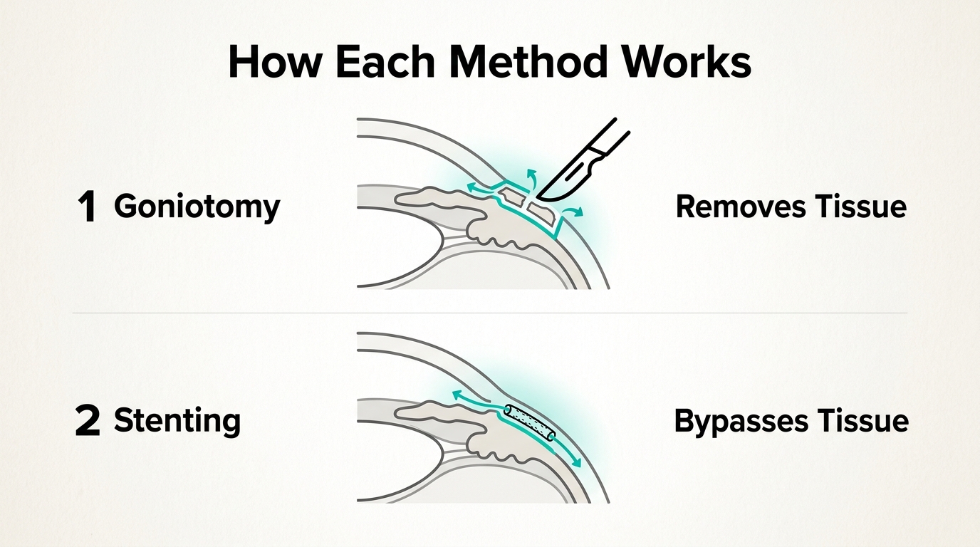
Goniotomy and trabecular micro-bypass stenting are two minimally invasive glaucoma surgeries (MIGS) that lower intraocular pressure by targeting the trabecular meshwork, the eye’s primary aqueous humor drainage structure. Each procedure uses a different mechanism: goniotomy excises or incises the tissue directly, while stenting implants a micro-device that bypasses it.
This guide covers how each procedure works, how their outcomes and risks compare, candidate selection for each approach, recovery and combined cataract surgery options, and the cost and insurance factors that may influence surgical planning.
Goniotomy uses instruments like the Kahook Dual Blade or catheter-based GATT systems to remove portions of the trabecular meshwork, restoring natural outflow through Schlemm’s canal. Stenting relies on FDA-approved devices, specifically the iStent and Hydrus Microstent, to create a permanent bypass channel through the meshwork without removing tissue.
Clinical comparisons suggest meaningful differences in IOP reduction, medication burden, and long-term durability. Stenting carries robust 5-year data showing sustained pressure control and reduced medication dependence, while goniotomy’s broader tissue removal may offer advantages for patients who need more aggressive outflow restoration or who have experienced prior failed procedures.
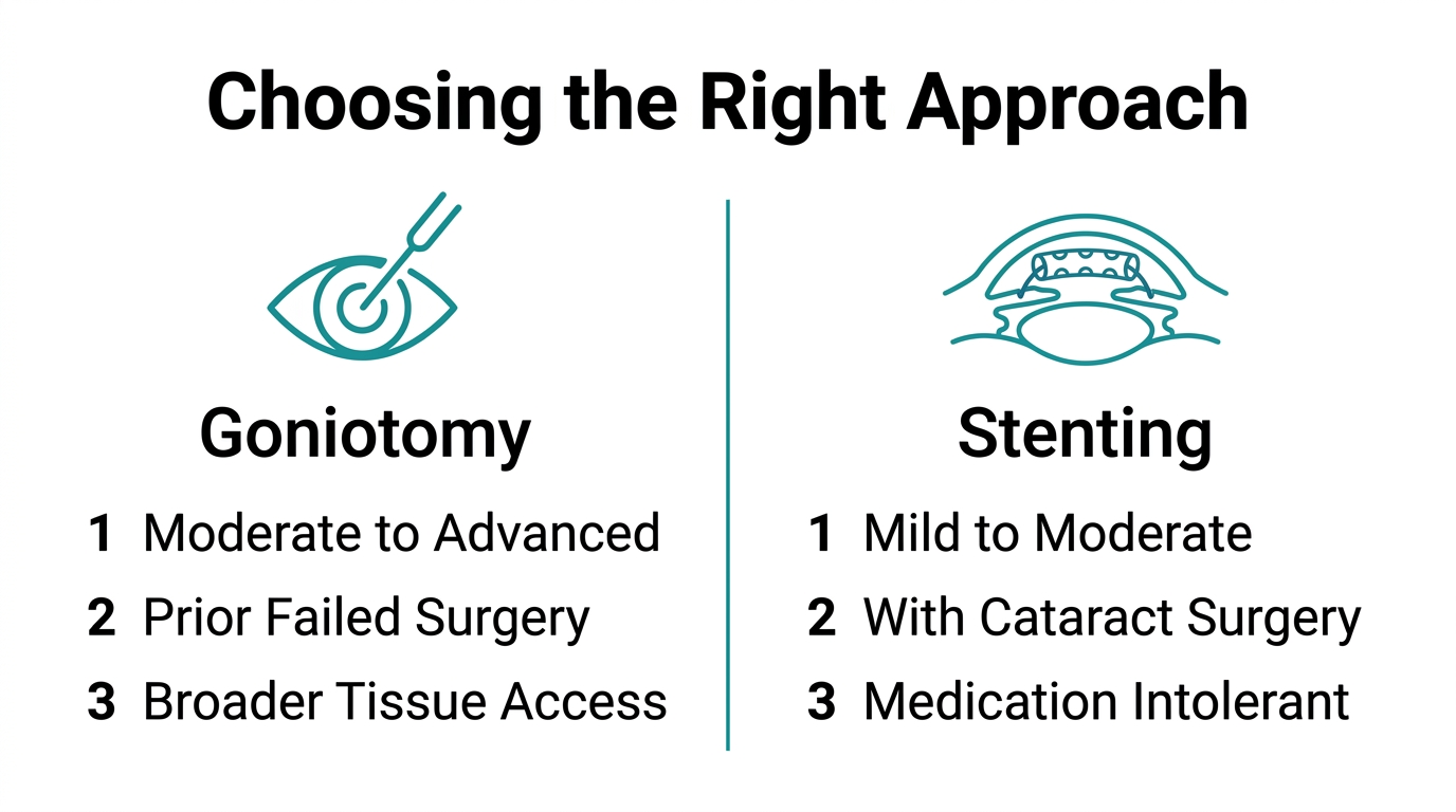
Candidate profiles differ as well. Stenting is primarily indicated for mild to moderate open-angle glaucoma in conjunction with cataract surgery, while goniotomy may extend to more complex cases including angle-closure presentations and revision scenarios. Complication profiles overlap but diverge in key areas; device malposition is the leading stenting concern, while hyphema is the most frequently observed event after goniotomy.
Costs, billing codes, and Medicare reimbursement rates also vary between the two approaches, with downstream medication savings potentially offsetting higher upfront procedural expenses for both.
What Is Goniotomy and How Does It Lower Eye Pressure?
Goniotomy is a minimally invasive glaucoma surgery (MIGS) that lowers intraocular pressure (IOP) by incising or removing tissue from the trabecular meshwork, the eye’s primary aqueous humor drainage structure. The following sections explain the procedure’s mechanism, the tools surgeons use, and how effectively it reduces IOP.
How Does Goniotomy Work to Reduce IOP?
Goniotomy works by cutting or excising the trabecular meshwork to restore aqueous humor outflow through Schlemm’s canal. When this drainage tissue becomes blocked or dysfunctional, IOP rises and damages the optic nerve. By removing the obstruction, goniotomy allows fluid to drain more freely, reducing pressure.
The extent of tissue removal varies by technique. According to a study published in Clinical Ophthalmology (Dove Medical Press), procedures like the Kahook Dual Blade (KDB) typically remove 90° to 120° of the trabecular meshwork, while approaches such as GATT or Trab360 perform a full 360° circumferential incision.
What Tools and Techniques Are Used in Goniotomy?
The tools used in goniotomy include the Kahook Dual Blade (KDB), the Goniotome, and catheter-based systems such as GATT (gonioscopy-assisted transluminal trabeculotomy) and Trab360. Each instrument targets the trabecular meshwork through the eye’s natural drainage angle using an ab interno approach, meaning no external incision is required. A gonioscopic lens provides the surgeon with a direct, magnified view of the drainage angle throughout the procedure.
Is Goniotomy an Effective Way to Lower Eye Pressure?
Goniotomy is an effective approach for lowering IOP, particularly in mild to moderate open-angle glaucoma. According to research published in Frontiers in Medicine, minimally invasive procedures including goniotomy are primarily indicated for mild and moderate open-angle glaucoma, especially in patients who do not tolerate topical medications. Broader 360° techniques, such as GATT, may offer greater pressure reduction than partial-arc excisions, making technique selection an important factor in surgical planning. For most patients, goniotomy offers a meaningful reduction in both IOP and medication burden with a favorable safety profile.
What Is Trabecular Micro-Bypass Stenting for Glaucoma?
Trabecular micro-bypass stenting is a minimally invasive glaucoma surgery (MIGS) that implants a tiny device into the trabecular meshwork to improve aqueous outflow and lower intraocular pressure (IOP). The following sections cover how these stents work, the two FDA-approved devices, and what candidates can expect.
How Does a Trabecular Micro-Bypass Stent Work?
A trabecular micro-bypass stent works by creating a direct channel through the trabecular meshwork, bypassing the primary site of aqueous outflow resistance and allowing fluid to drain into Schlemm’s canal more freely. This reduces the buildup of IOP without removing tissue. As Dr. Arsham Sheybani notes, these devices “are designed to help lower intraocular pressure by improving aqueous outflow through the trabecular meshwork.” Because the trabecular meshwork is a key site of outflow resistance in open-angle glaucoma, targeting it directly makes stenting one of the more mechanistically precise options available within the MIGS category.
What Are the FDA-Approved Trabecular Stents?
The two FDA-approved trabecular micro-bypass stents are the iStent and the Hydrus Microstent. The iStent Trabecular Micro-Bypass Stent received FDA approval on June 25, 2012, for mild to moderate open-angle glaucoma at the time of cataract surgery. The Hydrus Microstent received FDA approval in August 2018, also for use alongside cataract surgery in adult patients with mild to moderate open-angle glaucoma. Both devices are approved for combined use with cataract surgery, not as standalone implants in their originally approved indications.
What Does the Stent Implantation Procedure Involve?
Stent implantation involves inserting a microscale device into the trabecular meshwork through a small corneal incision using a goniolens for visualization. The procedure is typically performed at the time of cataract surger. Device malpositioning is the most commonly reported complication with iStent and Hydrus implants in the United States, making precise angle visualization a critical step. Given how technique-dependent accurate placement is, surgeon experience with gonioscopy and angle anatomy plays a significant role in outcomes.
How Does Goniotomy Compare to Stenting for Glaucoma?
Goniotomy and stenting differ in their mechanisms, IOP-lowering magnitude, medication reduction, and surgical technique. The sections below compare these procedures across four key clinical dimensions.
How Does IOP Reduction Differ Between Goniotomy and Stenting?
IOP reduction differs between goniotomy and stenting primarily in scope and mechanism. Goniotomy removes trabecular meshwork tissue directly, with procedures like the Kahook Dual Blade (KDB) excising 90° to 120° of the TM, while GATT and Trab360 perform a full 360° circumferential incision. Stenting devices, by contrast, bypass the TM rather than remove it. According to a comparison published in Clinical Ophthalmology (Dove Medical Press), these structural differences translate into measurable differences in surgical outcomes between excisional goniotomy and stent-based approaches. Tissue removal in goniotomy may produce broader outflow access, making technique selection an important part of surgical planning.
How Do Success Rates Compare Over Time?
Success rates over time vary between goniotomy and stenting, with both approaches showing durability in the right patient populations. Patients commonly ask about long-term success and visual recovery speed when comparing MIGS procedures. This real-world concern reflects a genuine clinical question: stenting outcomes are well-documented through pivotal trials, while longer-term goniotomy data continues to evolve. For patients weighing these options, follow-up frequency and baseline glaucoma severity are important factors in predicting durable success.
How Does Medication Reduction Differ After Each Procedure?
Medication reduction differs meaningfully between goniotomy and stenting, with stenting supported by robust long-term data. iStent inject produced significant and durable 5-year reductions, including nearly a 10-mmHg IOP decrease and a reduction of nearly 2 medications, in both standalone and combined phacoemulsification use, according to Glaukos clinical data. Comparable 5-year medication reduction data for goniotomy remains more limited in published literature. From a practical standpoint, patients who rely heavily on topical medications and experience tolerance issues may find stenting’s documented medication-sparing effect particularly valuable in shared surgical decision-making.
How Do Operative Times and Techniques Compare?
Operative times and techniques compare notably between goniotomy and stenting, reflecting their different surgical philosophies. Stenting procedures involve inserting a micro-device into Schlemm’s canal through the trabecular meshwork, often during combined cataract surgery. Goniotomy requires precise gonioscopic visualization and controlled tissue excision, particularly for 360° approaches like GATT. Both procedures are performed ab interno through a small corneal incision, avoiding external bleb formation. The technique choice often reflects surgeon experience and the patient’s specific drainage anatomy as much as any fixed time advantage.
Who Is a Good Candidate for Goniotomy?
Good candidates for goniotomy include patients with open-angle glaucoma, angle-closure glaucoma, and those who have experienced prior failed procedures. The following sections cover which patient profiles benefit most from this approach.
Who May Benefit Most from Goniotomy with Open-Angle Glaucoma?
Patients who may benefit most from goniotomy with open-angle glaucoma are those with mild to moderate disease who need meaningful IOP reduction without a permanent implanted device. Goniotomy works by directly excising or incising the trabecular meshwork, restoring aqueous outflow through the eye’s natural drainage anatomy rather than bypassing it with a stent.
For patients with advanced angle-closure glaucoma, a 2025 study published in JAMA Ophthalmology found that phacogoniotomy and phacotrabeculectomy both provided sustained surgical success with no additional complications at two years. This positions goniotomy as a viable option even in more complex anatomical presentations, not just straightforward open-angle cases.
Can Goniotomy Help Patients with Prior Failed Procedures?
Yes, goniotomy can help patients with prior failed glaucoma procedures, though the risk profile differs from primary cases. According to a study published in the Journal of Glaucoma, observed complications in eyes with failed prior surgery include hyphema (13.2%), transient IOP spikes (7.9%), and corneal edema (5.2%). For moderate-to-advanced cases, procedures like the XEN Gel Stent or trabeculectomy may be more appropriate than trabecular bypass stents.
Dr. Steven R. Sarkisian Jr. emphasizes that “achieving the best visual outcome with the fastest visual recovery should always be the first priority, even in patients with glaucoma,” which supports careful patient selection rather than defaulting to goniotomy in all revision scenarios.
Who Is a Good Candidate for Trabecular Micro-Bypass Stenting?
Good candidates for trabecular micro-bypass stenting are adults with mild to moderate open-angle glaucoma, particularly those undergoing concurrent cataract surgery. The following sections cover the ideal glaucoma severity profile and how combined cataract surgery candidacy affects stenting decisions.
Who May Benefit Most from Stenting with Mild to Moderate Glaucoma?
Patients who may benefit most from stenting are those with mild to moderate open-angle glaucoma, particularly when they do not tolerate topical medications. Both FDA-approved trabecular stents target this population specifically: the iStent received FDA approval on June 25, 2012, and the Hydrus Microstent received FDA approval in August 2018, each indicated for mild to moderate open-angle glaucoma in conjunction with cataract surgery. According to Frontiers in Medicine, stenting and goniotomy are primarily indicated for mild and moderate cases where topical treatment is poorly tolerated.
Patients with more advanced disease may not achieve sufficient IOP control through trabecular bypass alone and should be evaluated for alternative procedures. Device malpositioning is the most commonly reported complication with stenting MIGS in the U.S., including the iStent and Hydrus, so anatomical suitability matters when selecting candidates.
Can Stenting Help Patients Undergoing Cataract Surgery?
Yes, stenting can help patients undergoing cataract surgery by allowing glaucoma management and lens replacement to be addressed in a single procedure. According to Johns Hopkins Medicine, most patients undergoing MIGS can resume daily activities such as reading, watching TV, or using electronic devices within the first few days following surgery, making the combined approach practical for eligible patients.
One financial consideration worth noting: Medicare reimbursement for trabecular meshwork stent placement was reduced in 2022 to just $34.26 above the reimbursement for cataract surgery alone, according to Ophthalmology Management. From a patient-empowerment standpoint, understanding this reimbursement gap is important because it may influence which combined procedure options a surgeon presents or prioritizes.
What Are the Potential Risks of Goniotomy?
The potential risks of goniotomy include intraoperative bleeding, transient pressure spikes, and corneal swelling. The following sections cover the most common complications and how frequently hyphema occurs after the procedure.
What Complications May Occur During or After Goniotomy?
The complications that may occur during or after goniotomy include hyphema, transient intraocular pressure (IOP) spikes, and corneal edema. According to a 2023 study published in the Journal of Glaucoma, these complications were observed in eyes with prior failed glaucoma surgery, where hyphema occurred in 13.2% of cases, transient IOP spikes in 7.9%, and corneal edema in 5.2%. While these events are generally short-lived and manageable, patients with compromised surgical histories may face a modestly elevated complication profile. Close postoperative monitoring is essential to detect and address these issues early.
How Common Is Hyphema After Goniotomy?
Hyphema is the most common complication after goniotomy, occurring in approximately 13.2% of cases in high-risk eyes, based on the same 2023 Journal of Glaucoma study. It results from bleeding into the anterior chamber when the trabecular meshwork and Schlemm’s canal are incised. In most patients, hyphema is self-limiting and clears without intervention within a few days. Surgeons generally consider this an acceptable and expected trade-off given goniotomy’s broader IOP-lowering mechanism.
What Are the Potential Risks of Stenting?
The potential risks of stenting include device malpositioning, obstruction, bleeding, and transient IOP elevation. The sections below cover complications that may arise during or after stent placement and how frequently malposition occurs.
What Complications May Occur During or After Stent Placement?
Complications that may occur during or after stent placement include device malpositioning, intraoperative hyphema, transient IOP spikes, and incomplete trabecular meshwork bypass. Because stents such as the iStent and Hydrus Microstent are implanted through the trabecular meshwork into Schlemm’s canal, precise placement is essential. Any deviation in angle positioning can reduce aqueous outflow and compromise IOP reduction. Inflammation and early postoperative pressure elevation may also occur, typically managed with topical medications during the first weeks after surgery.
How Common Is Stent Obstruction or Malposition?
Stent malposition is the most common complication associated with MIGS stenting devices in the United States, including the iStent and Hydrus Microstent, according to EyeWorld. Malposition can occur when the device is not correctly seated within Schlemm’s canal, either obstructing the lumen or sitting outside the intended drainage pathway. When obstruction or malposition is identified early, repositioning or additional intervention may restore function. Surgeons experienced in gonioscopy-guided implantation tend to achieve more consistent placement, making surgical technique a meaningful factor in reducing this risk.
What Should You Expect During Recovery from Each Procedure?
Recovery from goniotomy and stenting follows broadly similar timelines, as both are minimally invasive glaucoma procedures. The H3s below cover what to expect after each surgery, including activity restrictions, follow-up schedules, and how quickly normal vision typically returns.
What Does Recovery Look Like After Goniotomy?
Recovery after goniotomy is generally short, with most patients resuming light daily activities within a few days. According to Johns Hopkins Medicine, patients undergoing MIGS procedures can typically return to activities such as reading, watching TV, or using electronic devices within the first few days following surgery. Post-operative follow-up visits are typically scheduled at day one, week one, week three, and then monthly for up to 90 days. Temporary blurring, mild redness, or light sensitivity may occur in the early days. Strenuous activity and eye rubbing should be avoided until cleared by a surgeon.
What Does Recovery Look Like After Stenting?
Recovery after stenting follows a similarly brief course. Most patients can resume reading and screen use within days, and the small-incision approach of device implantation means tissue disruption is minimal. Post-operative visits typically mirror the MIGS schedule: day one, week one, week three, and monthly check-ins through 90 days. Because stents remain permanently in the trabecular meshwork, early follow-up appointments are particularly important for detecting device malposition before it affects IOP control.
Can Goniotomy or Stenting Be Combined with Cataract Surgery?
Yes, both goniotomy and stenting can be combined with cataract surgery, and doing so is one of the most common approaches in modern glaucoma management. Performing a MIGS procedure alongside phacoemulsification allows surgeons to address elevated intraocular pressure and lens opacity in a single operative session, reducing overall patient burden.
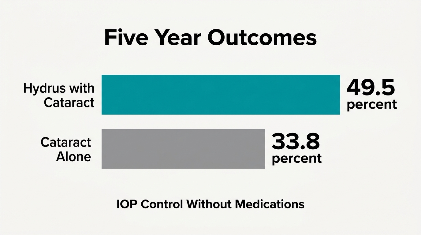
The long-term data supporting combined surgery is compelling. According to the HORIZON trial published in Ophthalmology, at 5 years, 49.5% of eyes receiving the Hydrus Microstent alongside cataract surgery achieved an IOP of 18 mmHg or less without medications, compared to 33.8% in the cataract surgery alone group (P = 0.003). Similarly, iStent inject has demonstrated significant and durable 5-year reductions in IOP of nearly 10 mmHg, with nearly a 2-medication reduction, in both standalone and combined phacoemulsification settings.
From a clinical standpoint, combining these procedures may be appropriate for patients with coexisting cataract and mild to moderate open-angle glaucoma. Rather than scheduling two separate surgeries with separate recovery periods, a single combined procedure can meaningfully reduce medication dependence while restoring visual clarity. For appropriate candidates, this represents may reduce the need for separate surgical sessions
How Do Costs and Insurance Coverage Compare?
Costs and insurance coverage for goniotomy versus stenting differ in billing codes, Medicare reimbursement rates, and long-term medication savings. The sections below cover CPT coding, Medicare reimbursement, and out-of-pocket cost considerations for each approach.
How Are Goniotomy and Stenting Billed for Insurance?
Goniotomy and stenting use different CPT billing codes, which affects reimbursement and insurance coverage decisions. Goniotomy is billed under CPT code 65820, an established Category I code with broad payer acceptance. Trabecular micro-bypass stents such as iStent and Hydrus were traditionally billed under Category III CPT code 0191T, a temporary code that some payers classify as investigational, potentially limiting reimbursement. This coding distinction is clinically significant: Category I codes generally face fewer prior authorization barriers than Category III codes, making goniotomy administratively simpler for many practices.
Does Medicare Cover Stenting Procedures Like Hydrus or iStent?
Medicare does cover trabecular micro-bypass stenting, but reimbursement rates have declined. According to Ophthalmology Management, Medicare reimbursement for trabecular meshwork stent placement was reduced in 2022 to only $34.26 more than reimbursement for cataract surgery alone. This narrow margin has led some surgeons and practices to reconsider the financial viability of stent-based MIGS relative to goniotomy, which carries a more favorable reimbursement structure under its established CPT code.
What Are the Long-Term Cost Implications for Patients?
The long-term cost implications of goniotomy versus stenting extend beyond surgical fees to include medication savings. A study conducted in Ontario, Canada found that combined cataract and iStent surgery cost Can$375 less than cataract surgery alone when accounting for long-term glaucoma medication savings. Similar savings logic applies to goniotomy, as both procedures aim to reduce or eliminate costly topical medications over time. For patients managing ongoing prescription costs, the downstream medication reduction may offset higher upfront procedural expenses, making surgical cost comparisons most meaningful when viewed across a multi-year horizon.
What Do Recent Studies Say About Long-Term Outcomes?
Recent studies on long-term outcomes show that both goniotomy and trabecular micro-bypass stenting can deliver durable IOP control, with the strongest evidence coming from the Hydrus Microstent’s HORIZON trial data and iStent’s FDA-tracked approval history.
What Does the HORIZON Trial Reveal About 5-Year Hydrus Outcomes?
The HORIZON trial reveals that the Hydrus Microstent produces meaningful, sustained IOP control well beyond the immediate postoperative period. At 5 years, 49.5% of eyes in the Hydrus group achieved an IOP of 18 mmHg or less without medications, compared to 33.8% in the cataract surgery alone group (P = 0.003), according to a long-term analysis published in Ophthalmology (AAO Journal). The Hydrus Microstent received FDA approval in August 2018, providing a regulatory benchmark that reflects this evidence base. For patients who want to reduce glaucoma drops over the long term, this level of medication-free IOP control may make Hydrus worth discussing with a surgeon.
What Do FDA Approval Timelines Indicate About Stenting Evidence?
FDA approval timelines indicate that trabecular micro-bypass stents have accumulated substantial supporting clinical evidence over more than a decade. The iStent Trabecular Micro-Bypass Stent received FDA approval on June 25, 2012, for the management of mild to moderate open-angle glaucoma at the time of cataract surgery, per the U.S. Food and Drug Administration. The six-year gap between iStent and Hydrus approval reflects the additional randomized controlled trial data required for each device. Longer regulatory track records generally translate to larger real-world outcome datasets, which strengthens confidence in safety and efficacy projections for both devices.
How Should You Evaluate Glaucoma Surgery with Your Eye Surgeon?
Evaluating glaucoma surgery requires understanding how procedures differ in mechanism, cost, recovery, and long-term outcomes. The following H3s cover surgeon-reviewed resources for combined procedures and the key clinical takeaways from this comparison.
Can Surgeon-Reviewed Cataract Surgery Resources Help You Understand Combined Glaucoma Procedures?
Yes, cataract surgery resources can help you understand combined glaucoma procedures by explaining how lens extraction and pressure-lowering interventions work together. For patients with chronic angle-closure glaucoma, lens extraction alone may reduce the need for additional glaucoma surgery, according to a Cochrane Library review. When open-angle glaucoma is present, devices like iStent inject and Hydrus are designed to lower intraocular pressure by improving aqueous outflow through the trabecular meshwork, as Dr. Arsham Sheybani has noted. Understanding terms such as MIGS, trabecular meshwork bypass, and Schlemm’s canal scaffolding helps patients engage more meaningfully in surgical planning conversations. Combined cataract and iStent surgery was also found to be Can$375 less expensive than cataract surgery alone over the long term when accounting for medication savings, according to data from Ontario, Canada.
What Are the Key Takeaways About Goniotomy vs Stenting?
The key takeaways about goniotomy vs stenting are that each procedure suits a different patient profile, disease severity, and clinical goal. Goniotomy removes trabecular meshwork tissue to restore outflow and may be preferred for more advanced cases or patients with prior failed procedures. Trabecular micro-bypass stents such as iStent and Hydrus are FDA-approved for mild to moderate open-angle glaucoma, particularly when combined with cataract surgery. Neither procedure is universally superior; the best choice depends on glaucoma severity, angle anatomy, prior surgical history, and tolerance for topical medications. Discussing these factors with your surgeon, using educational resources, is the most reliable path to an informed decision.