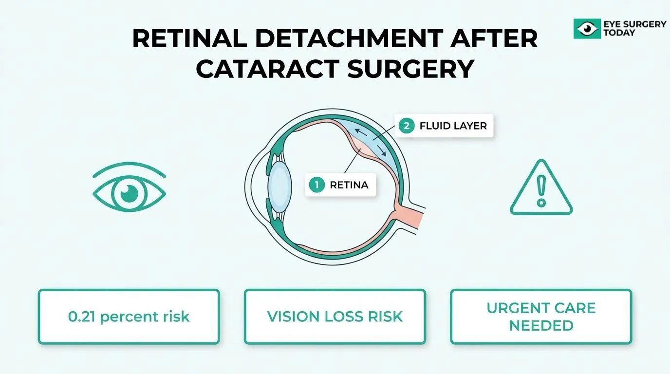
Retinal detachment after cataract surgery is a postoperative complication in which the neurosensory retina separates from the retinal pigment epithelium, potentially leading to permanent vision loss if left untreated.
This guide covers the clinical definition and causes of pseudophakic retinal detachment, warning symptoms and risk factors, diagnostic and treatment approaches, long-term vision outcomes, and prevention strategies.
Retinal detachment after cataract surgery occurs when vitreoretinal tractional forces create a retinal break, allowing liquefied vitreous to accumulate beneath the retina. Cataract surgery can accelerate this process, and complications such as posterior capsule rupture with vitreous loss may dramatically elevate risk.
The condition is uncommon, affecting roughly 1 in 500 cataract surgeries within the first year for patients over 40. However, risk may remain elevated for up to 20 years following the procedure, making long-term monitoring essential.
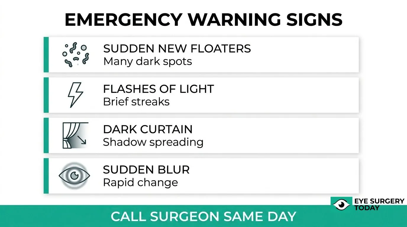
Sudden new floaters, flashes of light, a curtain or shadow spreading across vision, and rapid blurring are the primary warning signs. These symptoms constitute a medical emergency requiring same-day evaluation, as delays in treatment can allow detachment to reach the macula and significantly worsen visual outcomes.
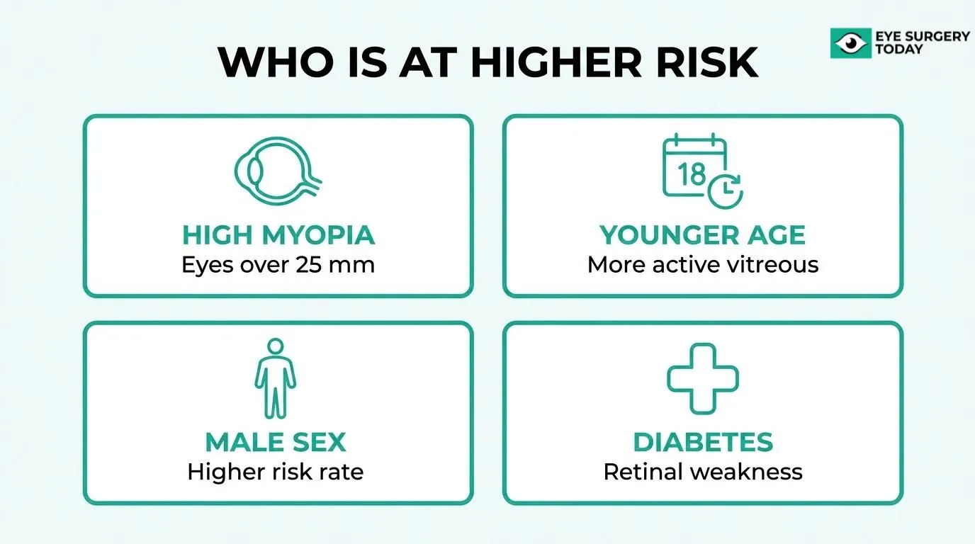
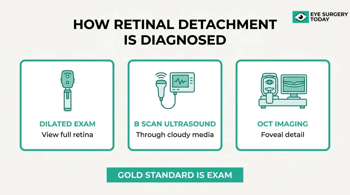
Several patient factors may increase susceptibility, including high myopia, prior retinal tears, younger age at surgery, and systemic conditions such as diabetes. Diagnosis relies on comprehensive dilated eye examination, sometimes supplemented by B-scan ultrasound or OCT imaging when direct visualization is limited.
Treatment ranges from laser photocoagulation or cryotherapy for contained tears to vitrectomy, scleral buckling, or pneumatic retinopexy for full detachment. Final reattachment rates can reach 93%, though visual recovery depends heavily on how quickly repair is performed and whether the macula was involved. Preoperative risk assessment, careful surgical technique, and consistent follow-up visits are among the most practical steps for reducing risk and protecting long-term vision.
How Is Retinal Detachment After Cataract Surgery Defined?
Retinal detachment after cataract surgery is a serious postoperative complication in which the neurosensory retina separates from the retinal pigment epithelium, threatening permanent vision loss if left untreated. The sections below cover the clinical definition, the mechanical process behind separation, and the types most relevant to cataract patients.
What Is Retinal Detachment After Cataract Surgery?
Retinal detachment after cataract surgery is a condition in which the neurosensory retina separates from the underlying retinal pigment epithelium following lens removal. According to the American Academy of Ophthalmology, if untreated, permanent vision loss may occur. The separation disrupts the retina’s oxygen and nutrient supply, making prompt recognition essential. For patients who have recently undergone cataract surgery, any sudden visual change warrants immediate evaluation.
How Does the Retina Separate From the Retinal Pigment Epithelium?
The retina separates from the retinal pigment epithelium through a process driven by vitreoretinal tractional forces. These forces create a full-thickness retinal break, allowing liquefied vitreous gel to enter the subretinal space. Once fluid accumulates beneath the neurosensory retina, progressive separation occurs. Cataract surgery can accelerate vitreous liquefaction and posterior vitreous detachment, making the retina more vulnerable to this mechanical sequence.
What Are the Main Types of Retinal Detachment That Can Follow Cataract Surgery?
The main types of retinal detachment that can follow cataract surgery are rhegmatogenous, tractional, exudative, combined tractional-rhegmatogenous, and macular hole-associated detachment. Rhegmatogenous retinal detachment is by far the most common type seen after cataract surgery, arising directly from a retinal break. Tractional and exudative forms are less frequent but remain clinically significant in patients with underlying conditions such as proliferative diabetic retinopathy or inflammatory eye disease.
What Causes Retinal Detachment After Cataract Surgery?
Retinal detachment after cataract surgery can result from surgical complications, pre-existing eye conditions, postoperative trauma, and patient-specific risk factors. The following sections examine each cause in detail.
How Can Surgical Technique and Intraoperative Complications Lead to Retinal Detachment?
Surgical technique and intraoperative complications can lead to retinal detachment primarily through posterior capsule rupture (PCR) with vitreous loss. When the posterior capsule tears during phacoemulsification, vitreous gel can prolapse forward, creating traction on the peripheral retina that may ultimately cause a full-thickness retinal break.
According to a study published in the British Journal of Ophthalmology, eyes experiencing PCR with vitreous loss had a hazard ratio of 12.83 (95% CI 5.62 to 29.30; p<0.001) for retinal detachment compared to uncomplicated cases, while no retinal detachment events occurred in eyes with PCR without vitreous loss. That contrast is clinically significant: vitreous loss, not capsule rupture alone, appears to be the critical driver of surgical risk.
How Do Pre-Existing Eye Conditions Increase the Risk of Retinal Detachment?
Pre-existing eye conditions increase the risk of retinal detachment by compromising the structural integrity of the retina and vitreous before surgery even begins. Conditions such as lattice degeneration, prior retinal tears, and a history of uveitis create zones of retinal weakness that become more vulnerable once the eye’s anatomy is altered by cataract surgery.
According to the National Eye Institute, warning signs of retinal detachment include a sudden increase in floaters, flashes of light, and a curtain or shadow over the visual field. Because these symptoms overlap with normal post-surgical recovery changes, patients with pre-existing retinal conditions require heightened monitoring after cataract surgery to avoid delayed diagnosis.
How Do Postoperative Trauma or Falls Contribute to Retinal Detachment?
Postoperative trauma or falls contribute to retinal detachment by applying sudden mechanical force to an eye that is still healing from surgery. In the weeks following cataract surgery, the eye remains more vulnerable to blunt impact, which can stress vitreoretinal adhesion points and trigger a tractional or rhegmatogenous tear.
This risk is particularly relevant for older adults, who face a higher fall frequency and may have pre-existing peripheral retinal weakness. Even a seemingly minor impact, such as bumping the eye or a low-velocity fall, may generate enough force to initiate vitreous traction on susceptible retinal tissue in the early postoperative period.
Can High Myopia or Other Patient Factors Raise the Risk of Retinal Detachment?
High myopia and other patient factors can raise the risk of retinal detachment after cataract surgery through anatomical and biometric characteristics that increase retinal fragility. A study published in the British Journal of Ophthalmology found that eyes longer than 25 mm carried a hazard ratio of 3.98 (95% CI 1.93 to 8.20; p<0.001) for retinal detachment compared to eyes measuring 23 to 25 mm.
Beyond axial length, other contributing patient factors include:
- Male sex, which is consistently associated with higher pseudophakic retinal detachment rates
- Younger age at surgery, which correlates with a more active, adherent vitreous
- Prior retinal tears or lattice degeneration in the operative eye
- A history of retinal detachment in the fellow eye
Understanding a patient’s full risk profile before surgery supports more targeted preoperative counseling and postoperative surveillance planning.
How Common Is Retinal Detachment After Cataract Surgery?
Retinal detachment after cataract surgery is uncommon but well-documented. The following sections cover the overall baseline risk, how that risk evolves over time, and how case complexity can influence outcomes.
What Is the Overall Risk of Retinal Detachment After Modern Cataract Surgery?
The overall risk of retinal detachment after modern cataract surgery is low, affecting approximately 1 in 500 cataract surgeries in patients over 40 within the first year, according to data from the IRIS Registry. A meta-analysis published in the Journal of Cataract and Refractive Surgery, analyzing 11 studies across 65,117 eyes, found that Nd:YAG laser capsulotomy, a common post-surgical procedure for cloudy vision, was associated with a 57% relative increase in retinal detachment risk. For most uncomplicated cases without this secondary procedure, the absolute risk remains well under 1%.
How Does Retinal Detachment Risk Change Over Time After Surgery?
Retinal detachment risk after cataract surgery does not peak immediately but may remain elevated for up to 20 years. According to a study published in the Transactions of the American Ophthalmological Society, the cumulative probability of retinal detachment following phacoemulsification was 0.27% at 1 year, rising to 0.71% at 5 years, 1.23% at 10 years, and 1.79% at 20 years. This gradual accumulation reflects ongoing vitreous changes that may stress the retina long after the original surgery. Patients and clinicians should treat long-term follow-up as an ongoing priority, not just an early post-operative concern.
How Does Retinal Detachment Risk Differ Between Standard and Complex Cataract Cases?
Retinal detachment risk differs substantially between standard and complex cataract cases, with intraoperative complications being the most influential factor. A British Journal of Ophthalmology study found that posterior capsule rupture with vitreous loss carried a hazard ratio of 12.83 compared to uncomplicated surgery, while eyes longer than 25 mm carried a hazard ratio of 3.98. In complex cases where the ocular media is unclear, diagnostic imaging plays an important role: B-scan ultrasound can identify detachments when direct visualization is limited, and OCT is particularly useful for assessing foveal involvement in tractional or exudative presentations.
What Types of Retinal Detachment Can Occur After Cataract Surgery?
Three types of retinal detachment can occur after cataract surgery: rhegmatogenous, tractional, and exudative. Each type has a distinct mechanism and clinical profile, covered in the sections below.
What Is Rhegmatogenous Retinal Detachment After Cataract Surgery?
Rhegmatogenous retinal detachment is the most common type to occur after cataract surgery. It develops when vitreoretinal tractional forces create a full-thickness retinal break, allowing liquefied vitreous gel to pass through and accumulate beneath the neurosensory retina, separating it from the retinal pigment epithelium. According to the American Academy of Ophthalmology, this fluid-driven separation is the defining mechanism of rhegmatogenous detachment. Cataract surgery accelerates posterior vitreous detachment, which can generate the traction needed to produce such a break, particularly in eyes with pre-existing retinal vulnerabilities.
What Is Tractional Retinal Detachment After Cataract Surgery?
Tractional retinal detachment is a type in which fibrovascular membranes pull the retina away from the retinal pigment epithelium without a retinal break. Rather than fluid entering through a tear, the retina is mechanically lifted by scar tissue or proliferative tissue growing across its surface. This form is most commonly associated with underlying conditions such as proliferative diabetic retinopathy. OCT imaging can be particularly useful for identifying the tractional components and assessing foveal involvement in these cases.
What Is Exudative Retinal Detachment After Cataract Surgery?
Exudative retinal detachment is a type driven by fluid accumulation beneath the retina from abnormal vascular leakage rather than from a retinal break or membrane traction. Inflammatory responses, choroidal pathology, or vascular disease can trigger subretinal fluid buildup after surgery. Because no retinal break is present, the mechanism and management differ substantially from rhegmatogenous detachment. OCT may help clinicians identify small amounts of subretinal exudate and confirm the exudative origin, which guides appropriate treatment planning.
What Symptoms of Retinal Detachment After Cataract Surgery Should You Watch For?
The symptoms of retinal detachment after cataract surgery include new floaters, flashes of light, a dark curtain or shadow across vision, and sudden blurring or distortion. Recognizing these warning signs early can be the difference between preserved sight and permanent vision loss.
How Do New Floaters Signal Possible Retinal Detachment?
New floaters signal possible retinal detachment when they appear suddenly and in greater numbers than before, rather than the occasional spots many people experience normally. According to the National Eye Institute, a sudden increase in floaters, described as small dark spots or squiggly lines drifting across vision, is one of the primary warning symptoms of retinal detachment.
After cataract surgery, the vitreous gel may shift or pull away from the retina, dislodging pigment cells or causing microscopic tears. Those displaced cells appear as a shower of new floaters. A single new floater is not always alarming, but a sudden burst warrants urgent evaluation by an eye care professional.
How Do Flashes of Light Warn About Retinal Detachment?
Flashes of light warn about retinal detachment by indicating mechanical traction on the retina. When the vitreous pulls or tugs against retinal tissue, photoreceptor cells misfire and the brain interprets these signals as brief streaks or arcs of light, typically in the peripheral field of one eye.
These flashes often appear in dim lighting and may persist for weeks. Unlike the flashes associated with migraine aura, retinal traction flashes do not involve headache and occur in a single eye. The National Eye Institute identifies flashes of light in one or both eyes as a key symptom requiring prompt attention.
What Does a Dark Curtain or Shadow in Vision Mean After Cataract Surgery?
A dark curtain or shadow in vision after cataract surgery means that the retina has likely detached in a specific region, blocking the visual field corresponding to that area. As detached retinal tissue loses its connection to the underlying retinal pigment epithelium, the affected zone stops transmitting visual signals, producing a fixed, spreading shadow rather than a transient blur.
This shadow may begin at the periphery and advance toward central vision. It does not fluctuate with blinking or eye movement, distinguishing it from temporary optical disturbances. This symptom represents a genuine ocular emergency and requires same-day evaluation.
How Can Sudden Blurring or Distortion of Vision Indicate Retinal Detachment?
Sudden blurring or distortion of vision can indicate retinal detachment when it develops rapidly, affects a distinct region of the visual field, and is not explained by typical post-surgical fluctuation. Distortion, where straight lines appear wavy or bent, may suggest that fluid has accumulated beneath the macula.
Unlike the gradual blur of residual refractive error after cataract surgery, detachment-related blurring tends to worsen quickly and may accompany other warning signs such as floaters or flashes. Any sudden, unexplained change in visual clarity following cataract surgery warrants immediate contact with your eye surgeon rather than a wait-and-see approach.
Who Is at Higher Risk for Retinal Detachment After Cataract Surgery?
Several patient-specific factors can meaningfully raise the risk of retinal detachment after cataract surgery. The H3 sections below cover four key risk categories: high myopia, prior retinal tears or detachment, age and sex, and systemic conditions such as diabetes.
How Does High Myopia Affect Retinal Detachment Risk After Cataract Surgery?
High myopia affects retinal detachment risk after cataract surgery by significantly increasing the likelihood of vitreoretinal complications. Eyes with axial length greater than 25 mm carry nearly four times the risk of retinal detachment compared to eyes measuring 23 to 25 mm, according to a study published in the British Journal of Ophthalmology (hazard ratio 3.98; 95% CI 1.93 to 8.20). Longer eyes have a thinner, more peripherally stressed retina, making them structurally more susceptible to tears once the vitreous shifts after lens removal. In clinical practice, high myopia is one of the most consistently predictive risk factors surgeons should identify and document before proceeding with cataract surgery.
How Do Previous Retinal Tears or Detachment Influence Future Risk?
Previous retinal tears or detachment influence future risk by indicating pre-existing vitreoretinal weakness that cataract surgery can further aggravate. Eyes with a history of retinal breaks have compromised peripheral retinal integrity, and the vitreous changes triggered by phacoemulsification may reopen or extend these vulnerable areas. While the research provided does not supply a specific recurrence hazard ratio for this subgroup, clinical guidelines consistently classify prior retinal detachment as a major independent risk factor. Surgeons should conduct a thorough peripheral retinal examination before surgery and consider prophylactic treatment of any existing tears to reduce post-operative detachment risk.
How Do Age and Sex Impact Retinal Detachment Risk After Cataract Surgery?
Age and sex impact retinal detachment risk after cataract surgery through differences in vitreous aging and axial eye length. Younger patients, particularly men, carry a disproportionately higher risk because male eyes tend to be longer on average and vitreous liquefaction with traction occurs differently across age groups. Data from the IRIS Registry published in Ophthalmology Science showed that among over 3.1 million eyes, rhegmatogenous retinal detachment within one year occurred in approximately 1 in 500 cataract surgeries in patients older than 40 years, with age-related patterns evident across the cohort. Knowing a patient’s age and sex profile helps risk-stratify surgical timing and post-operative follow-up frequency.
How Do Systemic Conditions Like Diabetes Affect Retinal Detachment Risk?
Systemic conditions like diabetes affect retinal detachment risk primarily through their impact on retinal tissue integrity and vitreoretinal anatomy before surgery even begins. Diabetic retinopathy can produce tractional forces on the retina from fibrovascular proliferation, making the retina more prone to both tractional and rhegmatogenous detachment after any intraocular procedure disturbs the vitreous. Patients with poorly controlled diabetes or advanced proliferative diabetic retinopathy require careful pre-surgical retinal evaluation and may need stabilization of their retinal disease before cataract surgery is appropriate. Understanding which systemic factors elevate risk allows surgeons and patients to plan the safest possible surgical pathway.
How Is Retinal Detachment After Cataract Surgery Diagnosed?
Retinal detachment after cataract surgery is diagnosed through a comprehensive eye examination, supported by specialized instruments and imaging tests when needed. The sections below cover examination methods, how tears and holes are identified, and when advanced imaging such as OCT or ultrasound is used.
What Eye Examination Methods Help Detect Retinal Detachment?
Eye examination methods that help detect retinal detachment include dilated fundus examination, slit-lamp biomicroscopy, and indirect ophthalmoscopy. During a dilated exam, the ophthalmologist widens the pupil with eye drops to get a full view of the peripheral retina, where most post-cataract detachments originate. Slit-lamp examination allows high-magnification assessment of the vitreous and inner retinal surface. According to Prevent Blindness, a comprehensive eye exam is the only way to diagnose retinal tears, making timely scheduling critical for any patient experiencing new visual symptoms after cataract surgery.
How Are Retinal Tears and Holes Identified After Cataract Surgery?
Retinal tears and holes are identified after cataract surgery primarily through dilated indirect ophthalmoscopy combined with scleral depression, a technique that allows the examiner to visualize the far peripheral retina where breaks most commonly form. Contact lenses designed for fundus examination can also be used at the slit lamp to improve the view of specific retinal zones. Because small breaks may produce few or no symptoms initially, a thorough peripheral retinal inspection is essential for every post-cataract patient reporting new floaters or photopsia.
When Are Imaging Tests Like OCT or Ultrasound Used to Diagnose Retinal Detachment?
Imaging tests like OCT or ultrasound are used to diagnose retinal detachment when the standard examination view is limited or when subtle structural changes require closer evaluation. B-scan ultrasound is particularly useful when media opacity, such as vitreous hemorrhage, blocks a clear fundus view, as it can reliably detect a detached retina behind an opaque medium. OCT provides high-resolution cross-sectional imaging of the retinal layers and is especially helpful for assessing foveal involvement and identifying tractional or exudative detachments, though it is not considered the gold standard for diagnosing all detachment types. Together, these tools give clinicians a more complete picture when clinical examination alone is insufficient.
What Are the Possible Risks and Complications of Retinal Detachment After Cataract Surgery?
The possible risks and complications of retinal detachment after cataract surgery include permanent vision loss, surgical complications during repair, and significantly worsened outcomes when treatment is delayed. The sections below cover each of these risks in detail.
How Can Retinal Detachment Lead to Permanent Vision Loss?
Retinal detachment can lead to permanent vision loss by separating the neurosensory retina from the retinal pigment epithelium, cutting off the photoreceptors from their essential support layer. According to the American Academy of Ophthalmology, when liquefied vitreous gel enters the subretinal space through a full-thickness retinal break, this separation progresses, and if left untreated, permanent loss of vision may occur. The risk is greatest when detachment reaches the macula, the central zone responsible for fine detail and color vision. Once photoreceptor cells are deprived of nourishment for too long, the damage cannot be reversed, even with successful surgical reattachment.
What Complications Can Occur During or After Retinal Detachment Surgery?
The complications that can occur during or after retinal detachment surgery include retinal redetachment, new or iatrogenic retinal breaks, cataract development or progression, and choroidal detachment. A Cochrane systematic review found that pars plana vitrectomy (PPV) was associated with lower rates of redetachment compared to scleral buckling, though new iatrogenic breaks were observed only in the PPV group, while choroidal detachment was more common with scleral buckling. In pseudophakic patients, the choice of surgical approach carries distinct risk profiles, and selecting the right technique requires careful clinical judgment based on detachment extent and patient anatomy.
How Can Delayed Treatment Worsen the Outcome of Retinal Detachment?
Delayed treatment worsens the outcome of retinal detachment by allowing the separation to extend to or beyond the macula, converting a macula-on detachment to a macula-off detachment. Research published in Frontiers in Public Health found a primary reattachment rate of only 60%, with a final reattachment rate of 93%, and visual acuity improved from 20/286 to 20/105 after surgery, indicating that even successfully treated cases may not recover full vision. From a clinical perspective, the speed of intervention is one of the most controllable factors in determining visual outcome, making prompt reporting of symptoms critical for any post-cataract patient.
How Is Retinal Detachment After Cataract Surgery Treated?
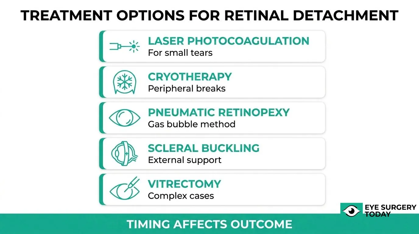
Retinal detachment after cataract surgery is treated using several surgical and procedural approaches, chosen based on the type, location, and severity of the detachment. The five main options are laser photocoagulation, cryotherapy, pneumatic retinopexy, scleral buckling, and vitrectomy.
When Is Laser Photocoagulation Used for Retinal Tears After Cataract Surgery?
Laser photocoagulation is used when a retinal tear is detected before full detachment has occurred. The procedure applies focused laser energy around the tear, creating small burns that form scar tissue, sealing the break and preventing fluid from entering the subretinal space. It is most effective for small, localized tears with no associated fluid accumulation. Because it is an in-office procedure requiring no incisions, ophthalmologists may recommend it as a first-line preventive intervention when a tear is caught early.
When Is Cryotherapy Used to Treat Retinal Breaks?
Cryotherapy is used to treat retinal breaks when laser photocoagulation is not feasible, such as when the break is located in the far peripheral retina or when media clarity is poor. The procedure applies a freezing probe to the outer surface of the eye, creating an adhesive scar around the break. Like laser treatment, it is most appropriate for contained breaks without extensive detachment. It can also be used in combination with other surgical techniques during more complex repairs.
When Is Pneumatic Retinopexy Recommended After Cataract Surgery?
Pneumatic retinopexy is recommended for select cases of rhegmatogenous retinal detachment where the break is located in the superior retina and the detachment is not too extensive. The procedure involves injecting a gas bubble into the vitreous cavity, which floats upward and tamponades the break, allowing fluid to absorb and the retina to reattach. It is typically combined with laser photocoagulation or cryotherapy to permanently seal the break. Patient positioning requirements after the injection are strict and essential to success.
When Is Scleral Buckling Surgery Used for Retinal Detachment?
Scleral buckling is used for rhegmatogenous retinal detachments, particularly in younger patients or those with detachments involving multiple breaks or significant vitreoretinal traction. The procedure involves placing a silicone band or element around the outside of the eye, indenting the scleral wall to relieve traction and close the retinal break. A Cochrane review found that, compared to vitrectomy, scleral buckling carried higher rates of choroidal detachment and a similar primary anatomical success rate, though evidence certainty was low. It remains a well-established option, especially when preserving the lens or managing peripheral breaks in younger pseudophakic patients.
When Is Vitrectomy Performed to Repair Retinal Detachment After Cataract Surgery?
Vitrectomy is performed when retinal detachment is complex, involves the posterior pole, or when other approaches are not suitable. The surgeon removes the vitreous gel and directly reattaches the retina using a combination of laser, gas tamponade, or silicone oil. Timing is critical: according to a study published in Eye, the risk of macula-on detachment converting to macula-off detachment is low (between 0.5% and 0.11%) when surgery is performed within 24 hours of presentation. Preserving macular attachment before surgery significantly improves visual outcomes, making prompt referral one of the most important factors in recovery.
What Can You Expect Before, During, and After Retinal Detachment Surgery?
Retinal detachment surgery involves three distinct phases: pre-surgical preparation, the repair procedure itself, and a recovery period that shapes final visual outcomes. The H3 sections below address what each phase typically involves.
How Should You Prepare If You Need Surgery for Retinal Detachment After Cataract Surgery?
Preparation for surgery for retinal detachment after cataract surgery typically involves a pre-operative evaluation, medication review, and fasting instructions from your surgical team. Because detachment can occur at any point following cataract surgery, understanding your personal risk timeline matters. According to a study published in the Transactions of the American Ophthalmological Society, the cumulative probability of retinal detachment after phacoemulsification rises from 0.27% at 1 year to 1.79% at 20 years, meaning risk persists well beyond the immediate postoperative window. Your ophthalmologist will assess which surgical approach is most appropriate before scheduling the procedure. Knowing this extended risk timeline is worth discussing with your surgeon before any cataract-related follow-up is completed.
What Typically Happens During Retinal Detachment Repair Procedures?
Retinal detachment repair procedures typically involve one or more surgical techniques, such as vitrectomy, scleral buckling, or pneumatic retinopexy, chosen based on the type, location, and extent of the detachment. Vitrectomy removes vitreous gel and allows direct access to the retina, while scleral buckling indents the eye wall to relieve traction. Pneumatic retinopexy uses a gas bubble to push the retina back into place. Each approach may be combined with laser photocoagulation or cryotherapy to seal the retinal break. The procedure is typically performed under local or general anesthesia and may take one to several hours depending on complexity.
What Is Recovery Like After Retinal Detachment Surgery?
Recovery after retinal detachment surgery varies by procedure type and may require face-down positioning, activity restrictions, and a period of reduced vision before improvement begins. Patients who undergo gas bubble tamponade are often instructed to maintain a specific head position for days to weeks to keep the bubble in contact with the repaired area. Air travel is generally restricted until the gas bubble fully absorbs. Swelling, mild discomfort, and temporary vision changes are common in the early weeks. Full recovery can take several months, and some patients require more than one procedure to achieve final reattachment.
How Soon After Retinal Detachment Treatment Might Vision Improve?
Vision improvement after retinal detachment treatment may begin within weeks of surgery, but final visual acuity can take months to stabilize. A 2022 study published in Frontiers in Public Health found that while the primary retinal reattachment rate was only 60%, the final reattachment rate reached 93%, with visual acuity improving from 20/286 to 20/105 over the full follow-up period. These results highlight that multiple procedures may be necessary and that patience is essential. Eyes with macular involvement typically recover more slowly and with less complete vision restoration than those where the macula remained attached throughout.
Can Retinal Detachment After Cataract Surgery Be Prevented or the Risk Reduced?
Retinal detachment after cataract surgery cannot always be prevented, but the risk can be meaningfully reduced through careful preoperative assessment, precise surgical technique, and consistent postoperative follow-up. The sections below cover what surgeons assess before operating, how intraoperative decisions affect risk, and what patients can do after surgery.
How Can Careful Preoperative Assessment Help Reduce Retinal Detachment Risk?
Careful preoperative assessment helps reduce retinal detachment risk by identifying high-risk anatomical and clinical features before surgery begins. Surgeons measure axial eye length, as eyes longer than 25 mm carry significantly elevated risk; according to a study published in the British Journal of Ophthalmology, eyes exceeding 25 mm had a hazard ratio of 3.98 for retinal detachment compared to eyes measuring 23 to 25 mm. Pre-existing peripheral retinal lattice degeneration, prior retinal tears, and a history of retinal detachment in the fellow eye are also documented during this evaluation. Identifying these factors allows the surgical team to plan accordingly, counsel the patient on elevated risk, and consider prophylactic retinal treatment before proceeding.
How Can Surgical Technique and Technology Influence Retinal Detachment Risk?
Surgical technique influences retinal detachment risk primarily through how well the posterior capsule is preserved during phacoemulsification. Posterior capsule rupture with vitreous loss is one of the most consequential intraoperative complications: the same British Journal of Ophthalmology study found a hazard ratio of 12.83 for retinal detachment when posterior capsule rupture occurred with vitreous loss, compared to no detachment events in eyes where rupture occurred without vitreous loss. Minimizing vitreous disturbance and maintaining capsular integrity are therefore central goals of modern phacoemulsification technique. Surgeons experienced with complex or high-myopia cases are better positioned to manage these intraoperative risks.
What Postoperative Behaviors May Lower the Risk of Retinal Detachment?
Postoperative behaviors that may lower retinal detachment risk include avoiding high-impact physical activity, contact sports, and any trauma to the eye during the early healing period. Patients should promptly report new floaters, flashes of light, or any shadow in their visual field rather than waiting for a scheduled appointment. Protecting the eye from accidental blows or falls reduces mechanical stress on the vitreoretinal interface while the eye stabilizes. Adhering to prescribed eye drop regimens supports controlled healing. These behaviors do not eliminate biological risk, but they reduce the chance of precipitating a tear in an already vulnerable retina.
How Do Regular Follow‑Up Visits Help Catch Retinal Problems Early?
Regular follow-up visits help catch retinal problems early by enabling dilated fundus examination at intervals when detachment risk is highest. According to Prevent Blindness, a comprehensive eye exam is the only way to diagnose retinal tears, and early detection allows treatment before detachment progresses. Retinal tears identified before fluid enters the subretinal space can often be sealed with laser photocoagulation or cryotherapy, avoiding the need for more complex surgery. Patients with known risk factors such as high myopia or prior retinal pathology benefit most from structured follow-up schedules. Consistent attendance at postoperative appointments is one of the most practical steps a patient can take to protect long-term vision outcomes.
When Should You Call Your Eye Surgeon About Possible Retinal Detachment After Cataract Surgery?
Knowing when to call your eye surgeon after cataract surgery can protect your vision. The sections below cover symptoms requiring emergency care, how quickly new floaters or flashes need attention, and which milder changes can wait for a scheduled visit.
What Sudden Vision Changes Require Immediate Emergency Care?
Sudden vision changes that require immediate emergency care include a new curtain or shadow spreading across your visual field, a sudden dramatic increase in floaters, or simultaneous flashes of light combined with any loss of peripheral vision. According to the National Eye Institute, retinal detachment is a medical emergency, and anyone experiencing these symptoms should contact their eye surgeon or go to an emergency room right away. Waiting even hours can allow detachment to extend beneath the macula, significantly worsening visual outcomes.
How Quickly Should You Be Seen If You Notice New Floaters or Flashes?
New floaters or flashes after cataract surgery warrant evaluation within 24 hours, not days. Research published in the journal Eye found that the risk of a macula-on detachment converting to a macula-off detachment was low (0.5 to 1.1%) when surgery was performed within 24 hours of presentation. Macular involvement is the single strongest predictor of poor long-term visual acuity, making same-day or next-day assessment essential. If your surgeon cannot see you urgently, an emergency eye clinic is appropriate.
Which Symptoms Can Safely Wait for a Scheduled Follow-Up Appointment?
Symptoms that can safely wait for a scheduled follow-up appointment include mild, stable blur that has not worsened since your last postoperative visit, a few pre-existing floaters that are unchanged in number or character, and minor light sensitivity without any new flashes or shadow. These findings are common after uncomplicated cataract surgery and are rarely signs of detachment. However, any symptom that changes, intensifies, or is accompanied by even a subtle shadow should prompt a same-day call to your surgeon rather than waiting. When in doubt, call first and let your care team decide the urgency.
How Should You Discuss Retinal Detachment Risks With Your Ophthalmologist Before Cataract Surgery?
Discussing retinal detachment risks with your ophthalmologist before cataract surgery involves asking targeted questions, reviewing your retinal history, and establishing a clear emergency plan. The H3s below cover each of these three preparation steps.
What Questions Should You Ask About Your Personal Retinal Detachment Risk?
The questions you should ask about your personal retinal detachment risk include your axial length, history of posterior capsule rupture risk, prior retinal tears, and whether Nd:YAG capsulotomy may be needed postoperatively. Axial length matters because, according to a study published in the British Journal of Ophthalmology, eyes longer than 25 mm carry nearly four times the retinal detachment hazard compared to eyes measuring 23 to 25 mm. Ask your surgeon to walk through each factor that applies to your eye specifically, then request a plain-language summary of your individual risk level so you can weigh it against the expected benefits of surgery.
How Can You Review Your Retina Health and Medical History Before Surgery?
You can review your retina health and medical history before surgery by requesting a dilated fundus examination and discussing any prior eye injuries, retinal tears, or family history of detachment at your preoperative appointment. According to Prevent Blindness, a comprehensive eye exam is the only way to diagnose retinal tears, and diagnostic instruments such as a slit lamp, contact lenses, and ultrasound may all be used during this evaluation. Bringing a written list of past eye conditions, systemic diagnoses, and previous surgeries helps your surgeon identify risk factors that imaging alone may not reveal.
How Should You Clarify the Warning Signs and Emergency Plan Before Surgery?
You should clarify the warning signs and emergency plan before surgery by asking your ophthalmologist to name the specific symptoms that require same-day emergency care and to confirm which clinic or emergency room to contact outside of office hours. According to the National Eye Institute, warning signs include a sudden increase in floaters, flashes of light, and a curtain or shadow crossing your visual field. Confirming the after-hours contact number, the nearest facility equipped for retinal repair, and how quickly you should seek care upon noticing symptoms gives you an actionable plan rather than uncertainty during a high-stress moment.
How Does Retinal Detachment After Cataract Surgery Affect Long‑Term Vision and Quality of Life?
Long-term vision outcomes after retinal detachment following cataract surgery depend on how quickly the detachment was treated and how much retinal tissue was involved. The H3s below cover timing and extent, daily activity impact, and rehabilitation options.
How Do Timing and Extent of Detachment Influence Long‑Term Vision?
Timing and extent of detachment directly influence how much vision a patient may retain after repair. When the macula remains attached at the time of surgery, visual prognosis is substantially better than in macula-off cases. Extent matters equally: a small peripheral detachment treated early carries a far better outlook than one involving the central retina.
According to a study published in Frontiers in Public Health, the final retinal reattachment rate reached 93%, and final Snellen visual acuity improved from 20/286 to 20/105 following repair, though primary reattachment was achieved in only 60% of cases. These figures illustrate that even after successful anatomical repair, meaningful visual deficits can persist, particularly when the detachment was extensive or involved the macula before treatment.
How Might Retinal Detachment Impact Daily Activities After Repair?
Retinal detachment may impact daily activities such as reading, driving, recognizing faces, and navigating low-light environments, even after anatomically successful repair. Persistent visual distortions, reduced contrast sensitivity, and central scotomas can make fine-detail tasks especially difficult.
For many patients, the gap between anatomical success and functional recovery is the most underestimated aspect of the condition. Vision that measures acceptably on a Snellen chart may still feel noticeably reduced during dynamic real-world tasks, and some patients report ongoing difficulty for months or years following surgery.
What Vision Rehabilitation or Low‑Vision Supports May Help After Retinal Damage?
Vision rehabilitation supports that may help after retinal damage include low-vision aids, orientation and mobility training, eccentric viewing therapy, and adaptive technology such as screen magnification software. These services are typically coordinated through a low-vision specialist or occupational therapist with experience in visual impairment.
Rehabilitation does not restore lost retinal tissue, but it can meaningfully improve a patient’s functional independence. Patients with residual central vision loss often benefit most from structured low-vision programs, particularly when rehabilitation begins early in the recovery period and is tailored to their specific visual field and acuity profile.
How Does Education From Trusted Eye‑Surgery Resources Help You Navigate Retinal Detachment Risks?
Education from trusted eye-surgery resources helps you navigate retinal detachment risks by translating complex clinical evidence into clear, actionable knowledge. The following sections explain how Eye Surgery Today supports safer decisions and summarize the core lessons from this article.
How Can Eye Surgery Today’s Cataract and Retinal Education Help You Make Safer Decisions?
Eye Surgery Today’s cataract and retinal education can help you make safer decisions by presenting surgeon-reviewed content that bridges the gap between clinical research and patient understanding. Knowing the warning signs, understanding your personal risk factors, and recognizing when to seek emergency care are decisions that depend entirely on having accurate information beforehand. Eye Surgery Today organizes this evidence clearly, without medical jargon, so patients can ask better questions, identify red-flag symptoms early, and approach consultations with confidence. Reliable education is often the difference between a patient who waits too long and one who seeks care in time to preserve vision.
What Are the Key Takeaways About Retinal Detachment After Cataract Surgery We Covered?
The key takeaways about retinal detachment after cataract surgery are focused on risk awareness, symptom recognition, and timely action. This article covered the following essential points:
- Retinal detachment is an uncommon but serious complication, with risk elevated by factors such as high myopia, posterior capsule rupture, and axial length greater than 25 mm.
- Warning symptoms, including sudden floaters, flashes of light, and a shadow or curtain across vision, require immediate emergency evaluation.
- Risk persists for up to 20 years after surgery, making long-term follow-up important.
- Treatment options range from laser photocoagulation for retinal tears to vitrectomy and scleral buckling for full detachment, with outcomes depending heavily on how quickly care is sought.
- Discussing your personal risk profile with your ophthalmologist before surgery is the most effective preventive step available.
Understanding these points puts you in the strongest position to protect your vision after cataract surgery.