Glaucoma is a group of optic neuropathies characterized by progressive retinal ganglion cell loss that can lead to irreversible blindness if undetected. Each type differs in its underlying mechanism, affected population, symptom presentation, and required treatment approach.
This guide covers the distinct glaucoma classifications, their symptoms and risk factors, how the condition is diagnosed, available treatment options, and how glaucoma relates to other eye conditions.
Open-angle glaucoma progresses silently as the trabecular meshwork loses drainage efficiency, while angle-closure glaucoma may present as a sudden emergency with severe pain and blurred vision. Less common forms, including normal-tension, congenital, pigmentary, exfoliation, neovascular, and traumatic glaucoma, each involve unique pathways that affect different age groups and demographics.
Symptoms range from completely absent in early open-angle disease to acute pain and nausea in angle-closure attacks, and risk factors span age, family history, ethnic heritage, elevated intraocular pressure, and systemic conditions such as diabetes.
Diagnosis relies on a coordinated evaluation that may include tonometry, gonioscopy, pachymetry, optical coherence tomography, and visual field testing to detect both structural and functional damage before significant vision loss occurs.
Treatment options range from prostaglandin analog eye drops and selective laser trabeculoplasty to traditional filtering surgery and minimally invasive glaucoma surgery (MIGS) devices, with the approach individualized based on disease severity and type.
Glaucoma also intersects with other eye conditions; cataract surgery may help lower intraocular pressure in angle-closure cases, while a glaucoma diagnosis can affect candidacy for refractive procedures like LASIK.
What Is Glaucoma and How Does It Affect Your Vision?
Glaucoma is a multifactorial optic degenerative neuropathy that progressively damages the optic nerve, leading to irreversible vision loss if left untreated. The sections below cover how glaucoma develops, how it damages vision, and how widespread it is globally.
How Does Glaucoma Damage the Optic Nerve?
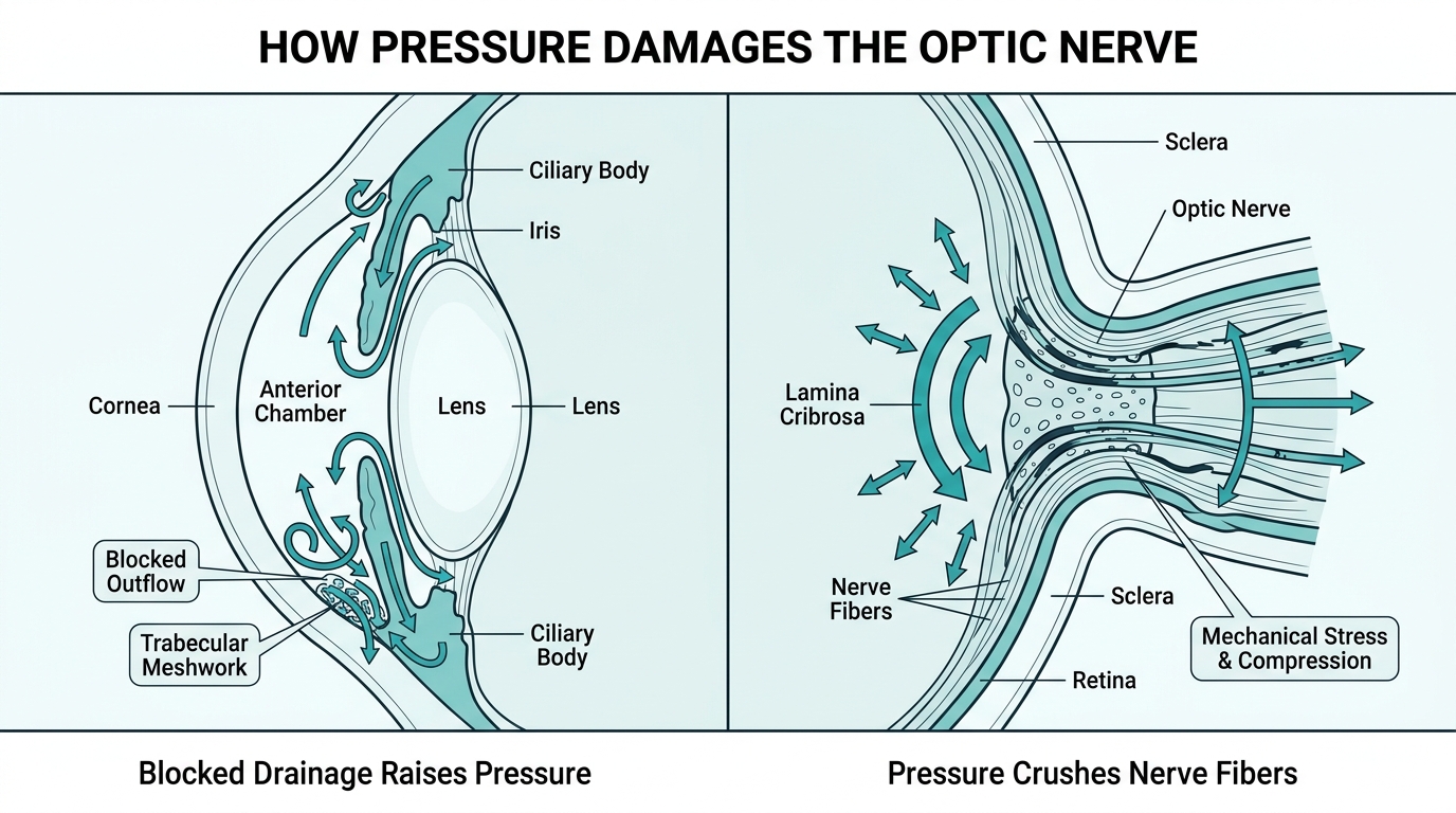
Glaucoma damages the optic nerve through the progressive loss of retinal ganglion cells, with elevated intraocular pressure (IOP) serving as a major modifiable risk factor. According to the CDC, elevated IOP causes mechanical stress on the lamina cribrosa, the structural plate through which optic nerve fibers exit the eye, ultimately disrupting axonal transport and triggering cell death. The biological mechanism involves increased resistance to aqueous humor drainage through the trabecular meshwork and uveoscleral pathways, which raises IOP and sustains that structural pressure over time. Because this damage is cumulative and largely silent in early stages, significant nerve loss can occur before any functional symptoms appear.
How Common Is Glaucoma Worldwide?
Glaucoma is the second leading cause of blindness after cataracts, affecting an estimated 76 million people worldwide as of 2020, according to the National Center for Biotechnology Information. In the United States alone, approximately 4.22 million people, representing 1.62% of adults aged 18 and older, are affected, with prevalence disproportionately higher among those of African and Hispanic descent. These numbers make early detection particularly critical, since vision lost to glaucoma cannot be restored.
What Are the Main Types of Glaucoma?
The main types of glaucoma are open-angle, angle-closure, normal-tension, congenital, secondary, pigmentary, exfoliation, neovascular, and traumatic glaucoma. Each type differs in cause, onset, and affected population. The sections below cover each type’s defining characteristics, prevalence, and key clinical distinctions.
Open-Angle Glaucoma
Open-angle glaucoma is the most common form, progressing silently as the trabecular meshwork gradually loses drainage efficiency while the anterior chamber angle remains physically open. According to a 2022 study published in the Journal of Global Health, the global incidence of primary open-angle glaucoma (POAG) was estimated at 23.46 per 10,000 person-years among individuals aged 40–79, with the highest rates in Africa and low sociodemographic index regions. Because the condition is typically asymptomatic until advanced stages, significant optic nerve damage often occurs before any symptoms are noticed. Clinical guidelines recommend an initial intraocular pressure (IOP) reduction of at least 20% to 30% from baseline for newly diagnosed POAG patients.
Angle-Closure Glaucoma
Angle-closure glaucoma occurs when the iris blocks the trabecular meshwork, preventing aqueous humor from draining. According to StatPearls, primary angle-closure glaucoma (PACG) has a global prevalence of approximately 0.6%, rising to 1.1% in Asian populations, and is commonly triggered by pupillary block that pushes the iris anteriorly. Unlike open-angle glaucoma, the acute form presents with sudden severe eye pain, headache, nausea, and blurred vision with halos, requiring urgent intervention. This rapid onset makes angle-closure glaucoma disproportionately responsible for acute vision emergencies relative to its prevalence.
Normal-Tension Glaucoma
Normal-tension glaucoma is a form of open-angle glaucoma in which characteristic optic nerve damage and visual field loss occur despite intraocular pressure remaining within the statistically normal range (below 21 mmHg). The underlying mechanism is thought to involve vascular insufficiency and increased optic nerve susceptibility rather than elevated IOP alone. Because standard tonometry readings appear normal, this type is frequently underdiagnosed without thorough optic nerve imaging and visual field testing.
Congenital Glaucoma
Congenital glaucoma is a rare developmental condition in which the eye’s drainage angle forms abnormally before birth, obstructing aqueous outflow. According to a study published in JAMA Ophthalmology, the birth prevalence of primary congenital glaucoma (PCG) is approximately 1 in 68,254 births in Western populations, yet it accounts for 5% to 18% of childhood blindness in some regions. Characteristic signs include enlarged eyes (buphthalmos), corneal clouding, and excessive tearing in infants. Early surgical intervention is typically necessary to preserve vision.
Secondary Glaucoma
Secondary glaucoma is glaucoma caused by an identifiable underlying condition, such as diabetic retinopathy, central retinal vein occlusion, uveitis, or prolonged corticosteroid use, rather than occurring independently. Treatment must address both elevated IOP and the root cause. Neovascular glaucoma, one of the more aggressive secondary forms, is covered in its own section below.
Pigmentary Glaucoma
Pigmentary glaucoma is a secondary open-angle glaucoma in which pigment granules shed from the iris clog the trabecular meshwork, progressively reducing aqueous outflow. According to EyeWiki (American Academy of Ophthalmology), this condition arises from the rubbing of the posterior iris against lens zonules and predominantly affects myopic males in their 30s. Physical activity can temporarily spike IOP in affected individuals by dislodging additional pigment.
Exfoliation Glaucoma
Exfoliation glaucoma is a secondary open-angle glaucoma caused by pseudoexfoliation syndrome (XFS), an age-related systemic condition in which abnormal elastic fibers accumulate in the anterior segment and clog the trabecular meshwork. According to a study in the Journal of Clinical Medicine (MDPI), XFS is the most common identifiable cause of secondary open-angle glaucoma worldwide. IOP fluctuations in exfoliation glaucoma tend to be more pronounced and harder to control than in POAG, often requiring earlier escalation of treatment.
Neovascular Glaucoma
Neovascular glaucoma (NVG) is a secondary glaucoma in which abnormal new blood vessels grow over the trabecular meshwork and iris, obstructing drainage. According to a study published in Medicina (MDPI), NVG carries a population prevalence of 0.01% to 0.12% but is highly aggressive, typically arising from retinal ischemia associated with diabetic retinopathy or central retinal vein occlusion. Management requires simultaneously treating the underlying ischemic driver and reducing IOP, often with a combination of anti-VEGF agents, laser, and surgery.
Traumatic Glaucoma
Traumatic glaucoma is elevated IOP resulting from blunt or penetrating eye injury, which may damage the trabecular meshwork through hyphema or angle recession. According to StatPearls, approximately 5% to 10% of patients with angle recession eventually develop glaucoma, sometimes years after the original injury. Because the condition can present long after trauma, any significant eye injury warrants ongoing IOP monitoring over time.
What Are the Symptoms of Each Type of Glaucoma?
The symptoms of each type of glaucoma vary significantly, from no noticeable signs in early open-angle disease to sudden, severe pain in acute angle-closure attacks. The H3s below cover open-angle, angle-closure, normal-tension, congenital, and secondary glaucoma symptoms individually.
What Are the Symptoms of Open-Angle Glaucoma?
The symptoms of open-angle glaucoma are largely absent in the early and middle stages of the disease. According to the Mayo Clinic, open-angle glaucoma is typically asymptomatic until advanced stages, meaning most patients experience no pain, redness, or noticeable vision changes until significant optic nerve damage has already occurred. As the disease progresses, peripheral vision gradually deteriorates, often described as tunnel vision. Because the loss is so gradual, many patients do not notice it until central vision is also affected. This silent progression is what makes routine screening so critical for at-risk individuals.
What Are the Symptoms of Angle-Closure Glaucoma?
The symptoms of angle-closure glaucoma depend on whether the attack is acute or chronic. Acute angle-closure presents as a medical emergency, with sudden severe eye pain, headache, nausea, vomiting, and blurred vision accompanied by halos around lights. The eye may appear red and the pupil may be fixed and mid-dilated. Chronic angle-closure, by contrast, may progress with minimal symptoms, similar to open-angle glaucoma, making it harder to detect without gonioscopic examination.
What Are the Symptoms of Normal-Tension Glaucoma?
The symptoms of normal-tension glaucoma mirror those of open-angle glaucoma: the condition is typically asymptomatic until optic nerve damage is advanced. Peripheral visual field loss develops progressively despite intraocular pressure remaining within the statistically normal range (below 21 mmHg). Because IOP readings appear unremarkable on routine screening, this form is frequently overlooked until a comprehensive optic nerve evaluation reveals characteristic cupping or nerve fiber layer thinning.
What Are the Symptoms of Congenital Glaucoma?
The symptoms of congenital glaucoma appear at birth or within the first few years of life and are distinct from adult presentations. The three hallmark signs are epiphora (excessive tearing), photophobia (light sensitivity), and blepharospasm (involuntary eyelid spasm). An enlarged cornea (buphthalmos) is also a characteristic finding, caused by elevated intraocular pressure stretching the immature ocular tissues. Parents and caregivers are often the first to notice these signs, making early pediatric eye evaluation essential.
What Are the Symptoms of Secondary Glaucoma?
The symptoms of secondary glaucoma vary based on the underlying cause, but generally follow patterns of either open-angle or angle-closure disease. Neovascular glaucoma, arising from retinal ischemia in conditions such as diabetic retinopathy or central retinal vein occlusion, often presents with severe eye pain, redness, and rapid vision loss. Traumatic glaucoma may be asymptomatic for years after an injury before progressive peripheral vision loss signals elevated pressure. Recognizing that secondary glaucoma symptoms are often masked by or attributed to the primary condition is a common clinical challenge worth emphasizing to patients.
What Causes Glaucoma and Who Is Most at Risk?
Glaucoma develops through a combination of mechanical, vascular, and genetic factors. The following sub-sections cover intraocular pressure’s role in optic nerve damage, plus the genetic, demographic, and medical conditions that raise risk.
What Role Does Intraocular Pressure Play in Glaucoma?
Intraocular pressure plays a central role in glaucoma by damaging the optic nerve through sustained mechanical stress. The eye continuously produces aqueous humor via the ciliary body and drains it through the trabecular meshwork and uveoscleral pathways; when drainage resistance increases, IOP rises. According to the CDC, elevated IOP causes mechanical stress on the lamina cribrosa, progressively destroying retinal ganglion cells. Dr. Robert N. Weinreb of UC San Diego states that “reduction of intraocular pressure is the only proven method to treat the disease,” with treatment goals individualized by disease severity and life expectancy. Elevated IOP also explains why glaucoma is a relative contraindication for LASIK: the suction ring transiently spikes IOP, and post-operative corticosteroids may elevate it further.
What Are the Genetic and Demographic Risk Factors?
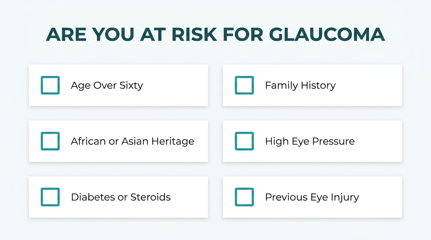
The genetic and demographic risk factors for glaucoma include advanced age, family history, and ethnic heritage. According to the National Eye Institute, individuals of African, Asian, or Hispanic descent face elevated risk, as do those with myopia, high IOP, or a history of long-term corticosteroid use. Family history is a particularly strong predictor, reflecting heritable variations in trabecular meshwork function and optic nerve susceptibility. In clinical practice, these overlapping risk factors mean that routine screening in high-risk demographic groups is one of the most practical tools for catching glaucoma before significant vision loss occurs.
Which Medical Conditions May Increase Glaucoma Risk?
Several medical conditions may increase glaucoma risk by directly elevating IOP or compromising optic nerve perfusion. Key conditions include:
- Diabetes mellitus, which is associated with neovascular glaucoma through retinal ischemia.
- Pseudoexfoliation syndrome, the most common cause of secondary open-angle glaucoma, involving abnormal elastic fiber deposits in the anterior segment.
- Primary angle-closure, where an enlarged crystalline lens crowds the drainage angle; cataract surgery may serve as a primary treatment by removing that lens.
- Long-term corticosteroid use, which can reduce trabecular outflow and raise IOP significantly.
Understanding these conditions helps clinicians identify patients who need earlier or more frequent glaucoma monitoring.
How Is Glaucoma Diagnosed?
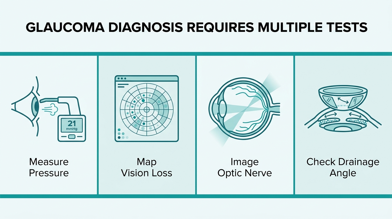
Glaucoma diagnosis relies on a multi-test evaluation that measures intraocular pressure, assesses the optic nerve, and maps functional vision loss. The following sections cover what a comprehensive exam involves, how IOP is measured, how visual field tests detect damage, and how optic nerve imaging confirms glaucomatous changes.
What Happens During a Comprehensive Glaucoma Eye Exam?
A comprehensive glaucoma eye exam involves several coordinated assessments rather than a single test. The eye care provider reviews patient history, measures visual acuity, examines the anterior chamber angle, assesses the optic nerve head, and evaluates central corneal thickness. Each component contributes a distinct data point that, when viewed together, allows clinicians to identify glaucoma at early, moderate, or advanced stages. No single finding alone confirms the diagnosis.
What Tests Help Measure Intraocular Pressure?
The tests used to measure intraocular pressure include tonometry and pachymetry, which are typically performed together. Applanation tonometry quantifies IOP in millimeters of mercury by measuring the force required to flatten a small area of the cornea. Pachymetry measures central corneal thickness because thinner corneas can cause tonometry to underestimate true IOP, while thicker corneas may overestimate it. Gonioscopy further complements these tests by visualizing the drainage angle to distinguish open-angle from angle-closure forms.
How Do Visual Field Tests Detect Glaucoma Damage?
Visual field tests detect glaucoma damage by mapping areas of reduced or absent vision across the patient’s full field of sight. According to NHS UK, glaucomatous damage typically manifests as arcuate scotomas or nasal steps that correspond directly to optic nerve head changes, making pattern recognition critical for staging disease severity. Automated perimetry presents light stimuli at controlled locations; the patient’s responses reveal which areas of the visual field have been compromised. Repeated testing over time tracks whether deficits are stable or progressing.
How Does Optic Nerve Imaging Help Diagnose Glaucoma?
Optic nerve imaging helps diagnose glaucoma by detecting structural loss in the retinal nerve fiber layer before functional deficits become apparent on visual field testing. Optical coherence tomography (OCT) generates high-resolution cross-sectional scans that quantify retinal nerve fiber layer thickness and identify thinning patterns consistent with glaucomatous neuropathy. According to the American Academy of Ophthalmology, a comprehensive glaucoma evaluation requires tonometry, gonioscopy, pachymetry, and OCT working together to build a complete diagnostic picture. Optic nerve photography also allows clinicians to document cup-to-disc ratio changes over serial visits, providing a visual record of progression. Given that glaucoma is a leading cause of irreversible vision loss, early structural detection through imaging can be important in diagnosis and monitoring.
What Are the Treatment Options for Glaucoma?
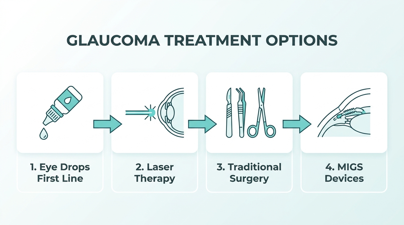
The treatment options for glaucoma include prescription eye drops, laser procedures, traditional surgery, and minimally invasive surgical techniques. The following sections cover how each approach works and what patients can expect.
How Do Prescription Eye Drops Help Manage Glaucoma?
Prescription eye drops help manage glaucoma by lowering intraocular pressure through several pharmacological mechanisms. Prostaglandin analogs, such as latanoprost and bimatoprost, are the most widely used first-line agents. According to a Cochrane Library review, prostaglandin analogs lower IOP by increasing uveoscleral outflow, typically achieving a 25% to 33% reduction from baseline. Other drop classes include beta-blockers, carbonic anhydrase inhibitors, and alpha-agonists, each working through distinct pathways to reduce aqueous humor production or improve drainage. For most newly diagnosed patients, eye drops remain the least invasive starting point before escalating to procedural options.
How Does Laser Treatment Work for Glaucoma?
Laser treatment for glaucoma works by improving the eye’s natural drainage to reduce intraocular pressure. Selective Laser Trabeculoplasty (SLT) is one of the most commonly used laser procedures, targeting pigmented trabecular meshwork cells without causing thermal damage to surrounding tissue, according to the Glaucoma Research Foundation. SLT can function as an initial therapy or an adjunct when eye drops alone are insufficient for open-angle glaucoma. Another option, laser peripheral iridotomy, addresses angle-closure glaucoma by creating a small opening in the iris to relieve pupillary block. In clinical practice, SLT may be used as an initial or adjunctive treatment option for open-angle glaucoma.
What Surgical Procedures May Treat Glaucoma?
The surgical procedures that may treat glaucoma include filtering surgery and tube shunt implantation, reserved for cases where drops and laser have not adequately controlled IOP. Trabeculectomy, according to the World Glaucoma Association, remains the gold standard for incisional glaucoma surgery, creating a new drainage pathway called a bleb. However, it carries recognized risks, including hypotony, bleb-related infections, and accelerated cataract formation. Tube shunt procedures, such as the Ahmed or Baerveldt implant, redirect aqueous humor through a silicone tube into an external reservoir and are often preferred in complex or refractory cases.
What Are Minimally Invasive Glaucoma Surgery Options?
Minimally invasive glaucoma surgery (MIGS) options include devices such as the Hydrus Microstent and iStent, which work by improving aqueous outflow through the trabecular meshwork with a significantly reduced risk profile compared to traditional filtering surgery. According to the British Journal of Ophthalmology, MIGS procedures offer a better safety profile than conventional incisional surgery and are frequently performed in combination with cataract surgery. This combination approach makes MIGS particularly attractive for patients who have both conditions, allowing surgeons to address elevated IOP and lens opacity in a single operative setting. For well-selected patients, MIGS may offer a less invasive surgical option with a different safety profile than traditional filtering surgery.
What Are the Potential Risks of Glaucoma Treatment?
The potential risks of glaucoma treatment vary by treatment type, ranging from mild medication side effects to serious surgical complications. The sections below cover the key risks associated with eye drops, laser therapy, and incisional surgery.
What Are the Side Effects of Glaucoma Eye Drops?
The side effects of glaucoma eye drops include local ocular reactions such as eye redness, stinging, blurred vision, and changes to eyelash growth or iris pigmentation. Prostaglandin analogs, including latanoprost and bimatoprost, are among the most widely used first-line agents, and while they effectively lower IOP by 25% to 33% according to a Cochrane Library review, they carry a recognized risk of periorbital fat atrophy and darkening of the iris over time. Systemic absorption can also cause cardiovascular or respiratory effects, particularly with beta-blocker drops such as timolol. Patient adherence tends to decline when side effects are poorly explained upfront, making clear counseling an underappreciated part of long-term glaucoma management.
What Are the Risks of Glaucoma Laser Surgery?
The risks of glaucoma laser surgery are generally mild and transient, making laser therapy one of the safer options available. Selective Laser Trabeculoplasty (SLT) targets pigmented trabecular meshwork cells without causing thermal damage to surrounding structures, which significantly limits its complication profile. Temporary IOP spikes, mild inflammation, and reduced efficacy over time are the most commonly reported outcomes. Because SLT preserves tissue architecture, it can typically be repeated, giving it a practical advantage over procedures with more permanent structural consequences.
What Are the Complications of Trabeculectomy?
The complications of trabeculectomy include hypotony, bleb-related infections, and accelerated cataract formation. According to the World Glaucoma Association, trabeculectomy remains the gold standard for incisional glaucoma surgery; however, the bleb it creates to divert aqueous humor is a persistent source of risk, as scarring or infection can lead to bleb failure or endophthalmitis. Hypotony, or abnormally low IOP, can cause choroidal effusion and vision loss if not managed promptly. For patients who require surgery but have moderate disease, Minimally Invasive Glaucoma Surgery (MIGS) devices such as the Hydrus Microstent and iStent offer a safer profile, though with less aggressive IOP reduction than trabeculectomy.
Can Glaucoma Be Prevented or Detected Early?
Glaucoma cannot be fully prevented, but early detection through routine screening can slow progression and preserve vision. The following sections cover risk reduction strategies and how regular eye exams identify glaucoma before significant damage occurs.
Can Glaucoma Be Prevented?
Glaucoma cannot be prevented entirely, as many risk factors such as age, genetics, and ethnicity are not modifiable. However, certain steps may help reduce risk or slow progression:
- Routine eye exams: Regular comprehensive screenings allow early IOP detection before optic nerve damage begins.
- IOP monitoring: Managing elevated intraocular pressure with prescribed drops may delay disease onset in high-risk individuals.
- Corticosteroid awareness: Limiting long-term corticosteroid use may reduce the risk of steroid-induced pressure elevation.
- Managing systemic conditions: Controlling conditions associated with secondary glaucoma, such as diabetic retinopathy, may reduce neovascular glaucoma risk.
According to the National Eye Institute, confirmed risk factors include advanced age, family history, African, Asian, or Hispanic heritage, high IOP, myopia, and long-term corticosteroid use. Identifying these risk factors early is, in practice, the most actionable form of “prevention” currently available.
How Is Glaucoma Detected Early?
Glaucoma is detected early through a comprehensive eye exam that measures intraocular pressure, evaluates the anterior chamber angle, and images the optic nerve. According to the American Academy of Ophthalmology, a complete glaucoma evaluation requires tonometry, gonioscopy, pachymetry, and optical coherence tomography (OCT) for retinal nerve fiber layer analysis. Visual field testing also detects early functional deficits, where damage typically appears as arcuate scotomas or nasal steps corresponding to optic nerve changes. Because open-angle glaucoma often produces no symptoms until advanced stages, these diagnostic tools are the only reliable way to catch the disease early, making routine screening genuinely life-changing for at-risk patients.
How Does Glaucoma Relate to Other Eye Conditions?
Glaucoma frequently overlaps with other eye conditions, both sharing risk factors and influencing treatment decisions. The following sections cover its relationship with cataracts and its impact on candidacy for other eye procedures.
What Is the Connection Between Glaucoma and Cataracts?
The connection between glaucoma and cataracts involves shared surgical pathways and direct treatment interactions. Cataract surgery may lower intraocular pressure in patients with primary angle-closure glaucoma, often serving as a primary treatment by removing the enlarged lens that contributes to angle crowding, according to a study published in The Lancet. Conversely, trabeculectomy, the gold-standard incisional glaucoma surgery, carries a known risk of accelerated cataract formation as a post-operative complication. Minimally Invasive Glaucoma Surgery (MIGS) procedures, such as the Hydrus Microstent or iStent, are frequently performed in conjunction with cataract surgery, offering a combined approach that addresses both conditions simultaneously.
Can Glaucoma Affect Candidacy for Other Eye Procedures?
Glaucoma can affect candidacy for refractive procedures such as LASIK. According to Review of Ophthalmology, glaucoma is a relative contraindication for LASIK due to the transient IOP spike caused by suction ring application during the procedure and the potential for steroid-induced pressure elevation post-operatively. Patients with glaucoma considering refractive surgery should discuss these risks with their eye care provider before proceeding. In practice, well-controlled glaucoma warrants careful case-by-case evaluation rather than automatic exclusion, making thorough pre-surgical screening essential.
How Can Eye Surgery Resources Help You Understand Glaucoma?
Surgeon-reviewed resources help you understand glaucoma by translating complex clinical evidence into clear, actionable knowledge. The following sections cover how Eye Surgery Today supports that goal and what key takeaways you should carry forward.
Can Eye Surgery Today Help You Learn About Glaucoma-Related Procedures?
Yes, Eye Surgery Today can help you learn about glaucoma-related procedures through educational content designed for patients navigating complex eye health decisions. The platform covers glaucoma types, diagnostic protocols, and treatment options, from laser therapy and eye drops to minimally invasive surgery, in accessible language free from unnecessary medical jargon. For patients weighing surgery options or trying to understand a recent diagnosis, clear educational information can support more informed conversations with a qualified ophthalmologist.
What Are the Key Takeaways About Glaucoma Types and Treatments?
The key takeaways about glaucoma types and treatments are that glaucoma is not a single disease but a spectrum of conditions, each with distinct causes, populations at risk, and management pathways. According to EyeWiki (American Academy of Ophthalmology), pigmentary glaucoma primarily affects males in their 30s, while primary angle-closure glaucoma is most prevalent in Asian populations. Primary congenital glaucoma, though rare in Western populations, accounts for 5% to 18% of childhood blindness in some regions, per JAMA Ophthalmology. Understanding these differences helps patients ask better questions and engage more meaningfully with their care team.