What is Glaucoma and Why May It Require Surgery?
Glaucoma is a group of eye conditions in which elevated intraocular pressure (IOP) may damage the optic nerve, leading to progressive, irreversible vision loss. The sections below explain how pressure harms the optic nerve and when medications are no longer sufficient to protect sight.
How Does Elevated Intraocular Pressure Damage Vision?
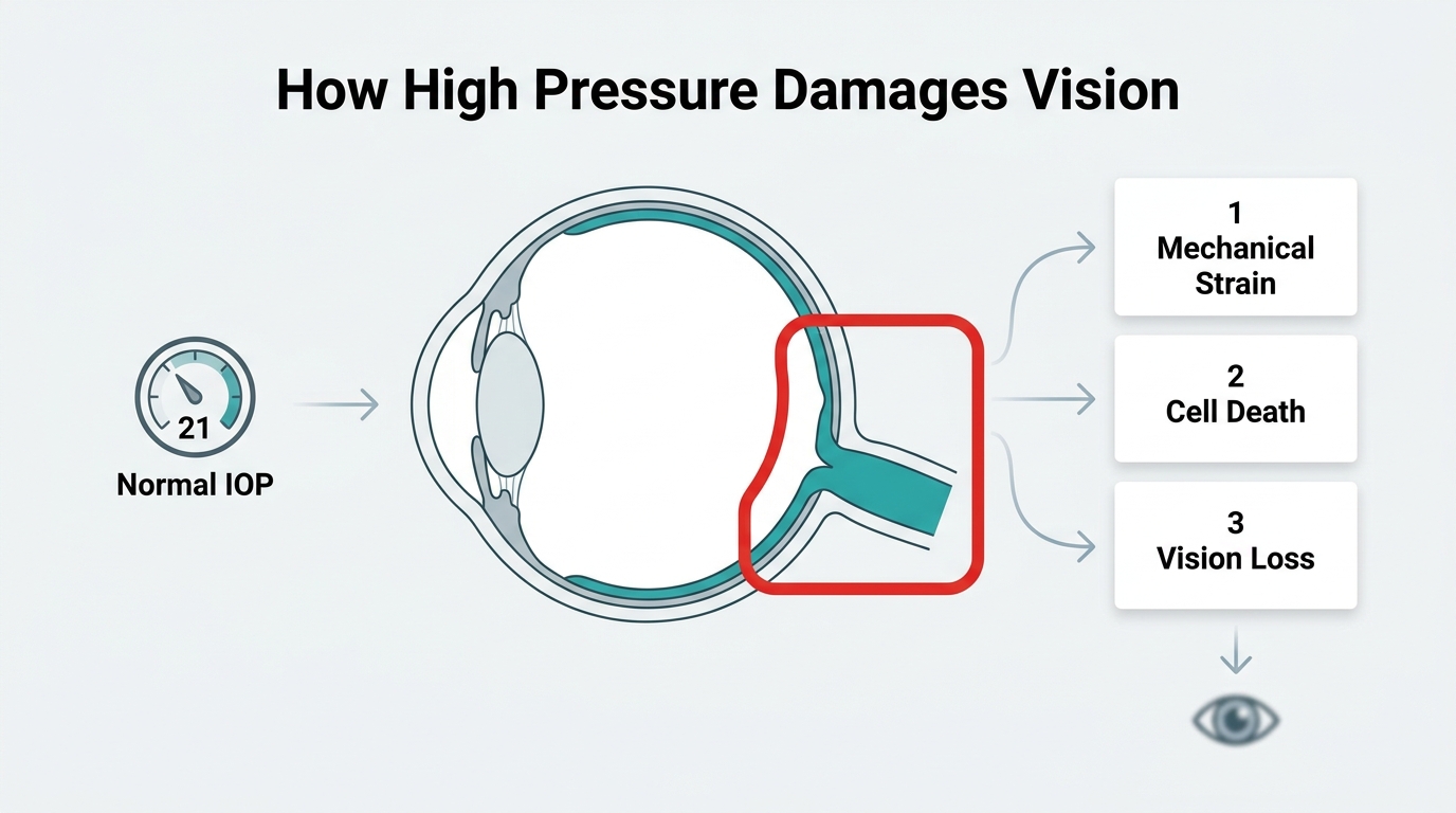
Elevated intraocular pressure damages vision by creating mechanical strain at the optic nerve head, disrupting axonal function and triggering retinal ganglion cell death. According to StatPearls (NCBI Bookshelf), intraocular pressure results from a balance between aqueous humor production by the ciliary body and its drainage through the trabecular meshwork and uveoscleral pathways. When this balance is disrupted, pressure rises. The American Academy of Ophthalmology’s EyeWiki defines the upper limit of normal IOP as approximately 21 mmHg, based on a Gaussian distribution of population measurements. Sustained pressure beyond this threshold may accelerate optic nerve injury even when visual symptoms are not yet apparent.
When Do Eye Drops or Medications Stop Being Enough?
Eye drops or medications stop being enough when they can no longer lower IOP to a target level that prevents further glaucomatous damage. According to a Cureus review, surgical intervention is typically considered when topical medications or laser treatments fail to achieve this protective threshold. At that point, a surgeon may recommend a procedure suited to the patient’s specific type and stage of glaucoma. Postoperative care then becomes essential: according to a National Institutes of Health (PMC) report, antibiotics and steroid eye drops may be required for up to 3 months after surgery to manage inflammation and reduce infection risk.
Who Is a Good Candidate for Glaucoma Surgery?
Surgical candidacy for glaucoma depends on the type of glaucoma, disease severity, and how well prior treatments have controlled intraocular pressure. The sections below cover candidacy for open-angle, angle-closure, secondary, and congenital glaucoma, plus when surgery may be considered first.
Who May Benefit from Surgery for Open-Angle Glaucoma?
Patients who may benefit from surgery for open-angle glaucoma are those whose IOP remains uncontrolled despite topical medications or laser therapy. Primary open-angle glaucoma is the most common form, and surgical options range from minimally invasive procedures to more traditional filtration surgeries, depending on disease stage. According to the European Glaucoma Society, surgical decision-making focuses on patients where target IOP is not achieved or progression continues despite medical therapy. Candidates typically have documented optic nerve damage or visual field loss that continues to advance despite maximum tolerated medical management.
Who May Benefit from Surgery for Angle-Closure Glaucoma?
Patients who may benefit from surgery for angle-closure glaucoma are those with a narrow or blocked drainage angle causing acute or chronic IOP elevation. Laser peripheral iridotomy is often the first intervention, creating a small opening in the iris to restore aqueous flow. When laser treatment fails to adequately control pressure, or if the angle remains functionally closed, additional surgical procedures may be necessary to prevent further optic nerve injury.
Who May Need Surgery for Secondary or Congenital Glaucoma?
Patients who may need surgery for secondary or congenital glaucoma are those whose elevated IOP results from an underlying condition, such as neovascular glaucoma, uveitic glaucoma, or a structural eye abnormality present from birth. These cases are often more complex and may require surgery earlier, since the underlying cause can make medical management less effective. Congenital glaucoma, in particular, frequently requires prompt surgical intervention because the developing visual system is especially vulnerable to pressure-related damage.
When Might a Doctor Recommend Surgery as a First Treatment?
A doctor might recommend surgery as a first treatment in cases of very high IOP at diagnosis, advanced optic nerve damage, or when medications are contraindicated or unlikely to achieve target pressure. However, according to updated Medicare policies, MIGS is not considered a first-line treatment for mild-to-moderate glaucoma and typically requires a trial of medical or laser therapy first. Surgery is most commonly considered primary when the severity of disease at presentation makes it clear that medications alone cannot adequately protect the optic nerve.
Understanding candidacy is the foundation for choosing the right surgical approach, which the next section covers in detail.
What Are the Types of Glaucoma Surgery?
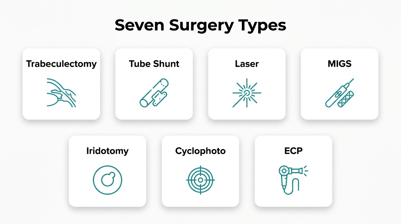
The types of glaucoma surgery include trabeculectomy, tube shunt implants, laser procedures, minimally invasive glaucoma surgery (MIGS), and cyclodestructive treatments. Each approach targets intraocular pressure (IOP) reduction through a different mechanism, and the right choice depends on glaucoma type, severity, and prior treatment history.
Trabeculectomy
Trabeculectomy is a filtering surgery in which a surgeon creates a small drainage channel in the sclera, allowing aqueous humor to bypass the blocked trabecular meshwork and collect beneath a protective tissue flap. This controlled outflow pathway reduces IOP. Trabeculectomy remains one of the most established surgical options for moderate-to-advanced open-angle glaucoma and is often enhanced with antimetabolites such as mitomycin C (MMC) to reduce scarring and improve long-term patency.
Tube Shunt Implant Surgery
Tube shunt implant surgery places a small silicone tube into the anterior chamber, routing aqueous humor to a plate implanted under the conjunctiva where it is absorbed. The TVT study found that tube shunt implantation achieved a higher rate of surgical success (defined as IOP ≤ 21 mmHg and reduced by ≥ 20%) than trabeculectomy with MMC after five years of follow-up. However, the PTVT study found that trabeculectomy with MMC achieved greater IOP reduction with fewer medications in eyes without prior incisional surgery, highlighting that neither procedure is universally superior.
Infection risk is a meaningful consideration for both approaches. According to a review published by Glaucoma Today, endophthalmitis rates are 2.0 per 1,000 surgeries for tube shunts and 1.5 per 1,000 for trabeculectomy, with infection rates across clinical settings ranging from 0.12% to 8.33% depending on follow-up duration.
Laser Trabeculoplasty
Laser trabeculoplasty uses focused laser energy applied to the trabecular meshwork to improve aqueous humor outflow and reduce IOP. Two primary forms are used clinically: selective laser trabeculoplasty (SLT) and argon laser trabeculoplasty (ALT). According to Ophthalmology Times Europe, SLT is reported to be equally effective as ALT in IOP lowering over a five-year period. SLT is commonly used as a laser trabeculoplasty option and has been reported to provide IOP lowering comparable to ALT over five years.
Laser Peripheral Iridotomy
Laser peripheral iridotomy (LPI) is a procedure in which a small hole is created in the peripheral iris using laser energy. This opening equalizes pressure between the posterior and anterior chambers, relieving pupillary block and widening the drainage angle. LPI is commonly used for angle-closure glaucoma because it addresses the underlying anatomical mechanism rather than lowering IOP through filtration.
Minimally Invasive Glaucoma Surgery (MIGS)
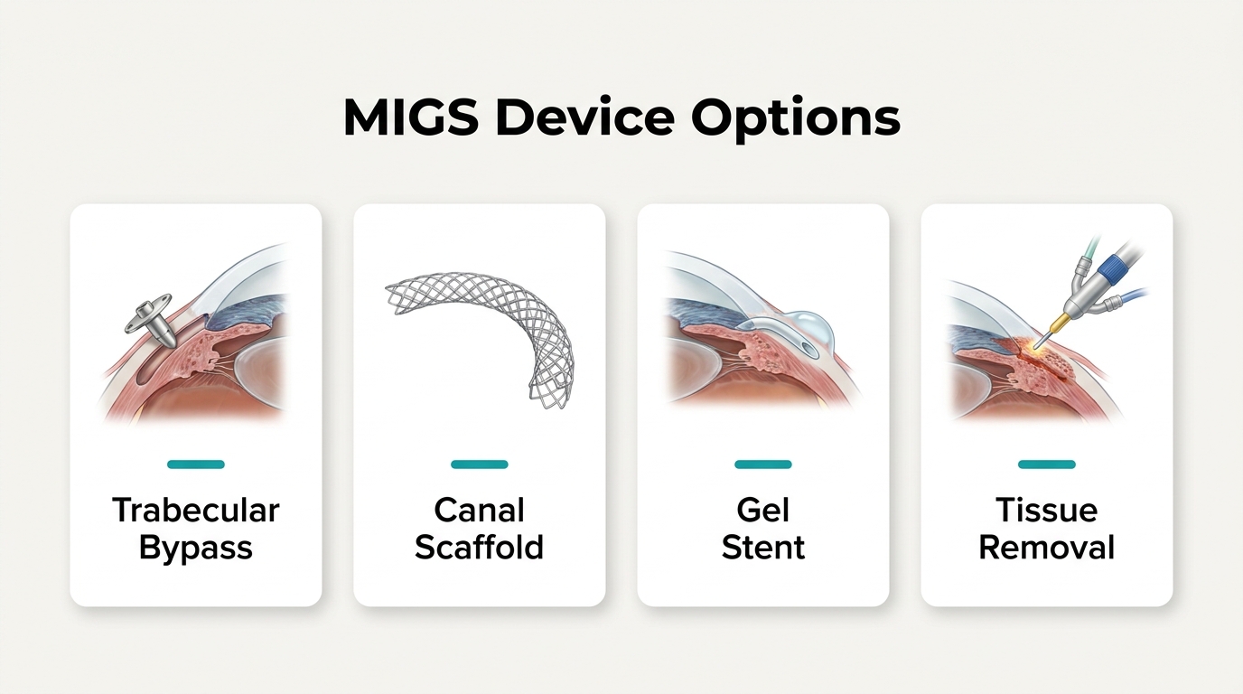
Minimally invasive glaucoma surgery (MIGS) describes a category of procedures that use micro-scale devices or techniques to enhance aqueous outflow through the eye’s natural drainage pathways with significantly less tissue disruption than traditional incisional surgery. According to EyeWiki, MIGS procedures offer a faster recovery profile than trabeculectomy due to smaller incisions and reduced surgical trauma. Common MIGS devices include the iStent, Hydrus Microstent, Xen Gel Stent, and Trabectome, each targeting a different drainage pathway.
Cyclophotocoagulation
Cyclophotocoagulation (CPC) is a cyclodestructive procedure that applies laser energy to the ciliary body to reduce aqueous humor production. Unlike filtering surgeries, CPC reduces IOP by decreasing fluid production rather than improving drainage. It is typically reserved for refractory glaucoma or eyes with poor visual potential where other surgical options have failed or are not appropriate, though evolving techniques have expanded its use in certain patient populations.
Endoscopic Cyclophotocoagulation (ECP)
Endoscopic cyclophotocoagulation (ECP) delivers laser energy directly to the ciliary processes under endoscopic visualization, allowing more precise tissue targeting than traditional transscleral CPC. According to Glaucoma Today, some studies report a mean IOP reduction of approximately 34% at 12 months postoperatively, a statistically significant result. ECP can be performed as a standalone procedure or combined with cataract surgery, and direct visualization allows the surgeon to target the ciliary processes during treatment.
With the full range of surgical types established, understanding which patients qualify for each procedure becomes the next critical step.
What Are the Different Minimally Invasive Glaucoma Surgeries?
The different minimally invasive glaucoma surgeries include iStent, iStent Inject, Hydrus Microstent, Xen Gel Stent, goniotomy, Trabectome, canaloplasty, and ab-interno canaloplasty. Each device or procedure targets a distinct anatomical pathway to reduce intraocular pressure with minimal tissue disruption.
iStent and iStent Inject
The iStent and iStent Inject are micro-bypass stents implanted into the trabecular meshwork to improve aqueous outflow. Both devices are among the most widely used MIGS options available, particularly for patients with mild-to-moderate open-angle glaucoma undergoing cataract surgery. Comparative data suggests nuanced performance differences: according to a 6-month subgroup analysis of 180 eyes reported by Dr. George Reiss at the 2025 AGS meeting, iStent may be slightly more efficacious in pseudophakic eyes and in SLT-naive eyes compared to Hydrus. However, the COMPARE trial found that the Hydrus Microstent outperformed two iStent devices as standalone treatment in reducing medication burden. Patient-specific factors, including prior laser history and lens status, appear to influence which device delivers the better outcome.
Hydrus Microstent
The Hydrus Microstent is an intracanalicular scaffold that spans approximately 8 mm of Schlemm’s canal, dilating the canal and bypassing the trabecular meshwork across multiple collector channel ostia. This broader canal engagement may explain its stronger performance in reducing medication dependence in head-to-head comparative data.
Xen Gel Stent
The Xen Gel Stent is a soft gelatin implant placed ab-interno to create a subconjunctival drainage channel, redirecting aqueous humor from the anterior chamber beneath the conjunctiva. It targets a bleb-forming pathway similar to trabeculectomy but through a less invasive internal approach, making it suitable for eyes with more advanced or refractory glaucoma.
Goniotomy and Trabectome Procedures
Goniotomy and Trabectome procedures both remove or ablate a portion of the trabecular meshwork to directly open aqueous outflow pathways. The Trabectome uses electrosurgical ablation of the trabecular meshwork and inner wall of Schlemm’s canal. According to the American Academy of Ophthalmology, standalone Trabectome surgery achieved a success rate of 46% at two years, while combined cataract and Trabectome surgery reached 85%, highlighting the meaningful benefit of pairing these approaches.
Canaloplasty and Ab-Interno Canaloplasty
Canaloplasty and ab-interno canaloplasty (ABiC) restore physiological aqueous outflow by dilating and viscodilating Schlemm’s canal and its collector channels without implanting a permanent device. The traditional external approach uses a suture placed under tension within the canal, while ABiC achieves similar canal dilation entirely from an internal incision with no suture. These procedures are particularly suited to patients seeking a device-free MIGS option with low bleb-related risk.
What Are the Potential Benefits of Glaucoma Surgery?
The potential benefits of glaucoma surgery include lowering intraocular pressure, reducing reliance on daily eye drops, and helping preserve remaining vision. The following H3s cover each of these outcomes in detail.
How May Glaucoma Surgery Help Lower Intraocular Pressure?
Glaucoma surgery may help lower intraocular pressure by creating new drainage pathways or reducing aqueous humor production, relieving mechanical strain on the optic nerve. According to JAMA Ophthalmology, glaucoma is a leading cause of irreversible blindness, and while surgery can lower IOP to prevent further damage, it cannot restore vision that has already been lost. This distinction matters enormously when setting patient expectations. The goal of surgical IOP reduction is preservation, not restoration, making timely intervention critical before significant nerve damage accumulates.
Can Glaucoma Surgery Reduce Dependence on Eye Drops?
Yes, glaucoma surgery can reduce dependence on eye drops for many patients. By improving aqueous outflow or suppressing its production, surgical procedures may achieve IOP control that previously required multiple daily medications. For patients managing complex drop regimens, reduced medication burden may help improve quality of life and eliminate concerns around adherence, side effects, and long-term medication costs. That said, some patients may still require drops after surgery, and outcomes vary by procedure type and disease severity.
How May Early Surgical Intervention Help Preserve Vision?
Early surgical intervention may help preserve vision by lowering IOP before irreversible optic nerve damage advances. Because glaucomatous vision loss cannot be recovered, intervening while functional vision remains intact offers the greatest protective benefit. Patients with rapidly progressing disease or those who have not responded adequately to medications may benefit most from earlier surgical consideration. From a clinical standpoint, waiting until vision loss becomes noticeable often means the opportunity for meaningful preservation has already narrowed significantly.
What Are the Possible Risks and Complications?
The possible risks and complications of glaucoma surgery range from short-term issues like inflammation and pressure fluctuations to longer-term concerns such as infection or device failure. The following sections cover short-term complications, long-term risks, and how risk profiles differ between MIGS and traditional procedures.
What Short-Term Complications Can Occur After Surgery?
Short-term complications that can occur after glaucoma surgery include elevated or low intraocular pressure, inflammation, bleeding inside the eye, and infection. Blurred vision and discomfort in the first days after surgery are also common. According to Glaucoma Today, preoperative assessment for ambulatory ophthalmic surgery includes evaluating sedation levels and ensuring adequate regional anesthesia to minimize sight-threatening complications. Careful surgical planning and close postoperative monitoring are important ways to catch and manage these issues early.
What Long-Term Risks Should Patients Understand?
Long-term risks patients should understand include scarring of the drainage pathway, device failure or migration, cataract formation, and persistent IOP fluctuations requiring additional treatment. Traditional procedures such as trabeculectomy carry a risk of bleb-related infection years after surgery, while tube shunt implants may require revision if the drainage tube becomes obstructed. These risks are manageable with regular follow-up but underscore why ongoing monitoring is essential after any glaucoma procedure.
How Does Complication Risk Differ Between MIGS and Traditional Surgery?
Complication risk differs meaningfully between MIGS and traditional surgery, with MIGS generally associated with a more favorable safety profile. According to EyeWiki, MIGS procedures have a faster recovery profile than traditional incisional surgeries like trabeculectomy due to smaller incisions and less tissue disruption. Traditional surgeries such as trabeculectomy carry higher rates of hypotony, bleb complications, and infection, while MIGS procedures generally involve smaller incisions and less tissue disruption. That said, MIGS typically achieves more modest IOP reductions, so the lower risk profile comes with a trade-off in pressure-lowering efficacy for advanced disease.
How Should You Prepare for Glaucoma Surgery?
Preparing for glaucoma surgery involves two key steps: completing required pre-operative evaluations and reviewing your current medications with your surgical team.
What Pre-Operative Tests and Evaluations Are Needed?
The pre-operative tests and evaluations needed for glaucoma surgery typically include a comprehensive eye examination, intraocular pressure measurement, visual field testing, optic nerve imaging, and a review of your overall medical history. Your surgical team will also assess your sedation needs and confirm that regional anesthesia can be safely administered. These evaluations help identify any conditions that could increase operative risk and ensure the chosen procedure is appropriate for your eye’s specific anatomy and disease stage.
Which Medications Should You Stop or Continue Before Surgery?
Your surgeon and anesthetist will review all medications you take before glaucoma surgery, including your current glaucoma eye drops. According to a study published by the National Institutes of Health (PMC), topical glaucoma medications can be systemically absorbed and may interact with anesthetic agents, which makes medication review an essential part of pre-operative planning. Blood thinners, anti-inflammatory drugs, and certain supplements may also need to be paused. Never stop or adjust any medication without explicit guidance from your care team, as some glaucoma drops must be continued right up to the day of surgery to maintain pressure control.
What Happens During a Glaucoma Procedure?
A glaucoma procedure varies in approach depending on the surgery type selected, but all share two common elements: anesthesia management and a defined operative timeframe. The sections below cover the anesthesia options typically used and how long patients can expect to be in the operating room.
What Type of Anesthesia Is Used for Glaucoma Surgery?
The type of anesthesia used for glaucoma surgery depends on the procedure’s complexity and the patient’s overall health. The anesthesia approach for glaucoma surgery depends on the procedure’s complexity, the patient’s health, and the surgical team’s assessment. For straightforward MIGS procedures such as iStent or Hydrus implantation, performed under CPT code 66991 alongside cataract surgery, topical or regional anesthesia is commonly sufficient. According to the European Glaucoma Society, surgical decision-making is individualized, particularly for patients who have not achieved target IOP through medical therapy, meaning anesthesia protocols must accommodate a range of procedure types and patient profiles.
How Long Does a Typical Glaucoma Procedure Take?
A typical glaucoma procedure varies depending on the surgical approach and whether glaucoma surgery is combined with another procedure, such as cataract surgery. MIGS procedures combined with cataract surgery are among the shortest, often completed in under 30 minutes. Traditional surgeries such as trabeculectomy or tube shunt implantation generally require more time due to greater tissue manipulation. According to Johns Hopkins Medicine, most patients notice a recovery in vision within days to weeks after surgery, though full stabilization may take longer depending on the specific procedure performed.
What Does Recovery After Glaucoma Surgery Look Like?
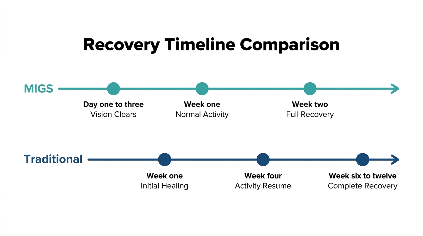
Recovery after glaucoma surgery varies by procedure type, spanning a few days for MIGS to several weeks for trabeculectomy. The sections below cover the first week, activity timelines, and required medications and follow-up visits.
What Can You Expect During the First Week of Recovery?
The first week of recovery after glaucoma surgery typically involves mild discomfort, blurred vision, and light sensitivity as the eye begins to heal. Most patients notice some vision improvement within days, though full stabilization may take longer depending on the procedure performed. According to Johns Hopkins Medicine, most patients notice a recovery in vision within days to weeks, with full stabilization varying by procedure type.
Common first-week experiences include:
- Mild redness, tearing, or a gritty sensation in the operated eye
- Temporary blurred or fluctuating vision
- Sensitivity to light and the need for protective eyewear
- Restrictions on bending, lifting, or straining
MIGS patients generally experience a shorter and less disruptive first week compared to those recovering from trabeculectomy or tube shunt surgery.
How Long Until Normal Activities Can Resume?
How long until normal activities can resume depends on the type of glaucoma surgery performed. MIGS procedures are considered to have a faster recovery profile than traditional incisional surgeries like trabeculectomy, due to smaller incisions and less tissue disruption, according to EyeWiki.
General return-to-activity timelines include:
- Driving: Typically cleared once vision stabilizes and the surgeon confirms adequate acuity, often within one to two weeks.
- Screen use and reading: Usually permitted within days for MIGS, but may take longer after trabeculectomy.
- Exercise and physical activity: Light activity may resume within one to two weeks; strenuous exercise and swimming are typically restricted for four to six weeks.
- Work: Desk-based work can often resume within a week; physical roles require longer recovery.
What Post-Operative Medications and Follow-Up Visits Are Needed?
Post-operative medications after glaucoma surgery typically include antibiotic and steroid eye drops to manage inflammation and reduce infection risk. According to a study published via the National Institutes of Health, these medications may be required for up to three months following surgery.
Key post-operative requirements include:
- Antibiotic eye drops: Used for several weeks to reduce infection risk, including serious complications such as endophthalmitis, which occurs at rates of 1.5 to 2.0 per 1,000 surgeries across trabeculectomy and tube shunt procedures, per Glaucoma Today.
- Steroid eye drops: Prescribed to control post-operative inflammation; dosing is typically tapered gradually over weeks.
- Follow-up visits: Scheduled by the surgeon based on the procedure type, healing progress, and IOP monitoring needs.
Patients with neovascular glaucoma may require closer monitoring, as research published in ScienceDirect indicates that surgical outcomes, including IOP control and visual prognosis, can be influenced by patient age at the time of surgery.
How Does Glaucoma Surgery Compare to Other Treatments?
Glaucoma treatment follows a stepwise approach, moving from eye drops to laser therapy to surgery as the condition progresses. The sections below compare surgery against medications and laser options across key clinical dimensions.
How Does Surgery Compare to Eye Drops for Lowering IOP?
Surgery may be considered for patients who cannot achieve target IOP through medication alone. Eye drops require daily adherence, and non-compliance is common; surgery delivers sustained pressure reduction without that burden. According to the European Glaucoma Society guidelines, surgical decision-making focuses specifically on patients where target IOP is not achieved or disease progresses despite medical therapy. For many patients, reaching the surgical threshold means eye drops have already proven insufficient, making the comparison somewhat self-selecting.
How Does Glaucoma Surgery Compare to Laser Treatments?
Glaucoma surgery generally achieves greater and more sustained IOP reduction than laser treatments, though laser carries a lower procedural risk. Selective laser trabeculoplasty may be equally effective as argon laser trabeculoplasty over five years, but neither typically produces the magnitude of pressure lowering achieved by trabeculectomy or tube shunts. Laser procedures are often used as an intermediate step before incisional surgery, particularly for mild-to-moderate glaucoma. Surgery remains the more definitive option when laser therapy fails to maintain IOP at a safe target.
Is Glaucoma Surgery Better Than Medication Long-Term?
Glaucoma surgery may offer better long-term IOP control than medication for patients with progressive disease. The HORIZON Study found that 73% of patients in the Hydrus group were medication-free at five years, compared to 48% in the cataract-only control group. Sustained medication-free control is clinically meaningful because it may reduce cumulative ocular surface side effects from long-term drop use. For patients managing multiple glaucoma medications with ongoing progression, surgery is often the more reliable long-term strategy.
How Do You Choose the Right Glaucoma Procedure?
Choosing the right glaucoma procedure depends on disease stage, overall eye health, and individual risk factors. The following sections cover how glaucoma severity and ocular health shape surgical decision-making.
How Does the Stage of Glaucoma Affect the Choice of Surgery?
The stage of glaucoma affects the choice of surgery by determining how aggressively intraocular pressure must be lowered and how much structural damage has already occurred. Elevated IOP creates mechanical strain at the optic nerve head, triggering extracellular matrix remodeling and retinal ganglion cell death, according to the National Institutes of Health. Mild-to-moderate cases often respond to less invasive options such as laser trabeculoplasty or MIGS devices. Advanced disease, where significant optic nerve damage has already progressed, may require procedures with greater IOP-lowering capacity, such as trabeculectomy or tube shunt implantation. According to the CDC, an estimated 4.22 million Americans were living with glaucoma in 2022, with 1.49 million experiencing vision-affecting disease, underscoring how often staging directly shapes treatment urgency. Matching procedure intensity to disease stage is an important factor in helping protect remaining vision.
What Role Does a Patient’s Overall Eye Health Play?
A patient’s overall eye health plays a critical role in determining which glaucoma procedure is appropriate and safe. Coexisting conditions such as cataracts, corneal disease, prior ocular surgeries, or a history of uveitis can all limit or favor certain surgical approaches. For example, a patient with concurrent cataract may benefit from a combined MIGS and cataract extraction procedure, while someone with severely compromised conjunctival tissue may not be a suitable candidate for trabeculectomy. Systemic health factors, including bleeding disorders or immunosuppression, also influence procedural risk. A thorough preoperative evaluation allows the surgical team to weigh each patient’s ocular and systemic profile against the available options, ensuring both safety and effectiveness are optimized for that individual.
How Should You Approach Glaucoma Surgery Alongside Cataract Care?
Combining glaucoma and cataract treatment into one procedure is a well-established strategy. The sections below cover how MIGS integrates with cataract surgery and summarize the key lessons from this article.
Can Cataract Surgery and MIGS Be Combined in One Procedure?
Yes, cataract surgery and MIGS can be combined in appropriate patients, and research supports measurable IOP benefit compared with cataract surgery alone. Canal-based devices like the iStent and Hydrus may lower IOP by 15 to 30 percent from baseline when implanted alongside cataract surgery. A meta-analysis published in Cureus found that MIGS combined with cataract surgery reduced IOP by a mean of 1.58 mmHg more than cataract surgery alone across pooled studies.
The efficiency advantage is also meaningful. Standalone Trabectome surgery achieved a success rate of 46% at two years, while combined cataract surgery plus Trabectome reached 85%, according to data published by the American Academy of Ophthalmology. Hypotony risk remains low, with reported rates of approximately 0.13%, though observed ranges vary across studies. For patients who are appropriate candidates for both procedures, combining them may deliver measurable IOP benefit compared with cataract surgery alone.
What Are the Key Takeaways About Glaucoma Surgery?
Glaucoma surgery encompasses a broad spectrum of options, from minimally invasive trabecular devices to traditional filtration surgery, each suited to different disease stages and patient profiles. Several practical conclusions emerge from this article:
- Surgical timing matters: intervention is typically considered when medications or laser therapy cannot achieve the target IOP needed to halt progression.
- MIGS procedures, particularly when combined with cataract surgery, offer meaningful IOP reduction with lower complication risk than traditional incisional surgery.
- Traditional approaches like trabeculectomy and tube shunts remain the standard for advanced or refractory glaucoma when MIGS is insufficient.
- Surgery lowers IOP to protect remaining vision but cannot restore sight already lost, making early intervention central to long-term outcomes.
- Recovery timelines and post-operative medication requirements vary by procedure, so patients benefit from understanding what to expect before surgery.
Eye Surgery Today provides educational content to help patients better understand glaucoma surgery options and related decisions.