Retina surgery is a group of specialized procedures that repair, preserve, or restore the delicate neural tissue lining the back of the eye. These operations address sight-threatening conditions through internal vitreous removal or external scleral reinforcement, distinguishing them from lens-based or corneal surgeries.
This guide covers the conditions that may require retina surgery, the main procedure types and how they work, candidacy and risk considerations, recovery timelines and post-operative care, and how retina surgery compares to other eye surgeries in scope and expected outcomes.
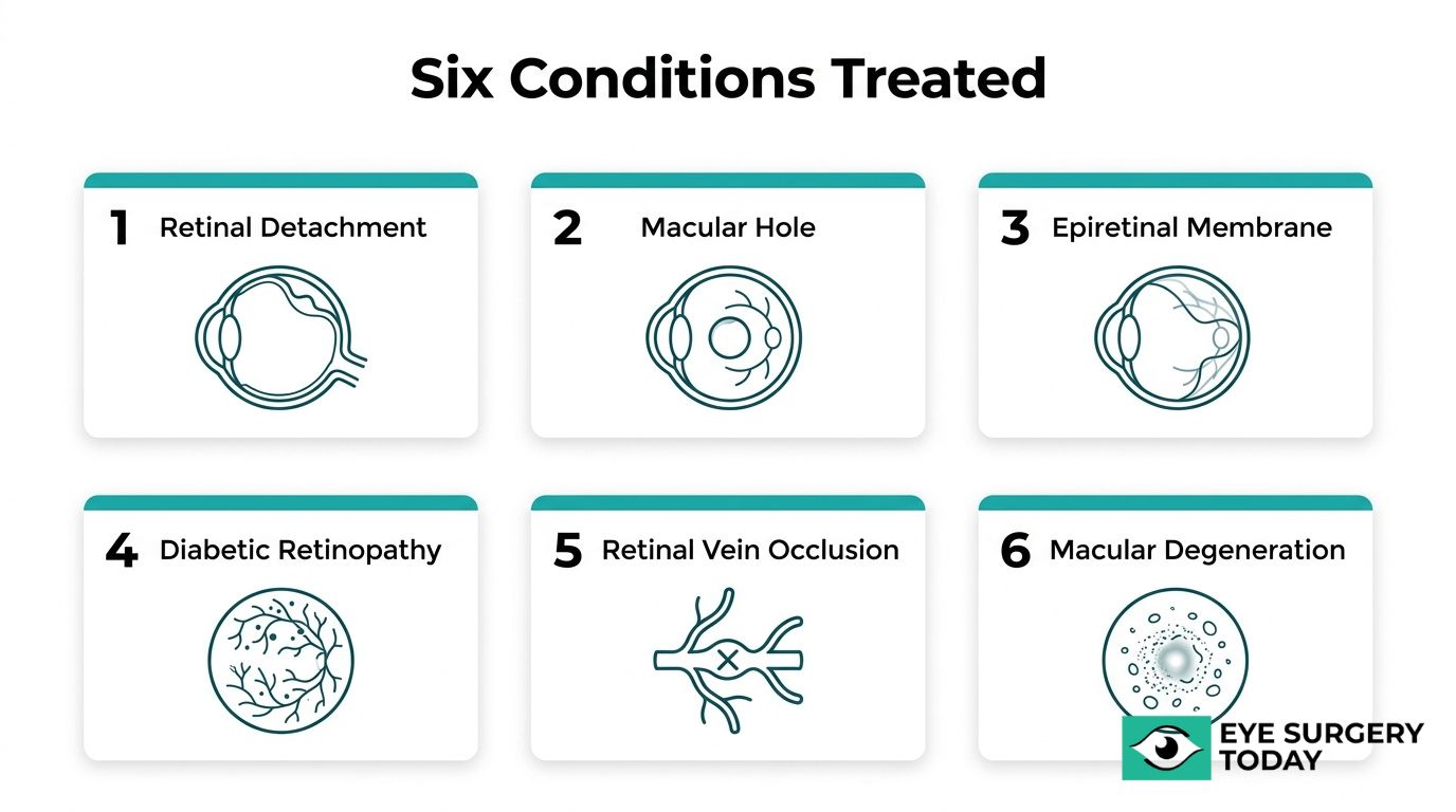
Retinal detachment, macular holes, epiretinal membranes, diabetic retinopathy, retinal vein occlusion, and macular degeneration each present distinct threats to vision. Some, like retinal detachment, demand urgent intervention, while others may be monitored before surgery becomes necessary.
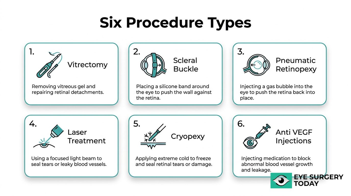
Procedures range from vitrectomy and scleral buckle surgery to pneumatic retinopexy, laser photocoagulation, cryopexy, and anti-VEGF intravitreal injections. Each technique targets a specific mechanism of retinal damage, and the recommended approach depends on the diagnosed condition and its severity.
Surgical candidacy involves factors such as macular involvement, timing of intervention, and overall ocular and systemic health. While serious complications remain uncommon, each procedure carries its own risk profile that patients should discuss with a retina specialist.
Recovery periods vary from days for laser photocoagulation to several weeks for vitrectomy, with full visual improvement often taking months. Strict post-operative positioning, prescribed eye drops, and activity restrictions may play a direct role in whether the surgical repair succeeds.
What Is Retina Surgery?
Retina surgery is a group of specialized surgical procedures designed to repair, preserve, or restore the delicate neural tissue lining the back of the eye. Unlike cataract surgery, which replaces a cloudy lens, or LASIK, which reshapes the cornea, retina surgery involves internal vitreous removal or external scleral reinforcement to treat conditions affecting the retina itself. These procedures address sight-threatening problems, such as retinal detachment, macular holes, epiretinal membranes, and complications from diabetic retinopathy.
A retinal specialist, also known as a vitreoretinal surgeon, performs these operations using microsurgical instruments and advanced imaging. Related terms for retina surgery include vitreoretinal surgery, pars plana vitrectomy, retinal reattachment, and macular hole repair. The specific procedure recommended depends on the diagnosed condition, its severity, and the patient’s overall eye health. Understanding what retina surgery involves can help patients feel more informed when discussing treatment options with their eye care provider.
What Eye Conditions May Require Retina Surgery?
Eye conditions that may require retina surgery include retinal detachment, macular hole, epiretinal membrane, diabetic retinopathy, retinal vein occlusion, and macular degeneration.
Retinal Detachment
Retinal detachment is a condition where the retina separates from the underlying supportive tissue at the back of the eye. Without prompt surgical intervention, permanent vision loss can result. According to a study published in JAMA Ophthalmology, rhegmatogenous retinal detachment carries an annual incidence of 6.3 to 17.9 per 100,000 population.
Surgical options for retinal detachment include vitrectomy, scleral buckle surgery, and pneumatic retinopexy. The specific approach depends on the type, location, and severity of the detachment. Early detection and timely repair can significantly improve visual outcomes, making retinal detachment one of the most time-sensitive conditions in all of ophthalmology.
Macular Hole
A macular hole is a small break in the macula, the central part of the retina responsible for sharp, detailed vision. This condition commonly affects people over the age of 55 and occurs most often in women, with a 5% to 15% risk of developing a hole in the fellow eye.
Vitrectomy is the standard surgical treatment. According to the American Society of Retina Specialists, vitrectomy surgery for macular holes has a success rate of over 90%, with patients regaining some or most of their lost vision. Given these outcomes, early evaluation is especially worthwhile for anyone noticing central vision distortion or missing spots in their field of view.
Epiretinal Membrane
Epiretinal membrane is a condition where a thin layer of fibrous tissue develops on the surface of the macula, causing visual distortion and blurriness. According to StatPearls, the mean age at diagnosis is 65 years, with an incidence in the first eye of 5.3%.
Not all epiretinal membranes require surgery. When the membrane significantly impairs vision or daily activities, vitrectomy with membrane peeling can improve visual clarity. Mild cases may simply be monitored over time, which makes regular retinal exams essential for tracking progression.
Diabetic Retinopathy
Diabetic retinopathy is damage to the retina’s blood vessels caused by chronic high blood sugar. According to the American Society of Retina Specialists, diabetic retinopathy develops in more than half of people who develop diabetes and is the most common cause of irreversible blindness in working-age Americans.
Treatment options range from anti-VEGF intravitreal injections to laser photocoagulation and vitrectomy, depending on disease stage. For proliferative diabetic retinopathy, anti-VEGF injections have shown more favorable visual acuity outcomes compared with panretinal photocoagulation at three years. This condition underscores why routine dilated eye exams are so critical for anyone living with diabetes.
Retinal Vein Occlusion
Retinal vein occlusion is a blockage of the veins that carry blood away from the retina, leading to swelling, hemorrhage, and potential vision loss. According to a study published in Retinal Physician, retinal vein occlusion prevalence ranges from 157 to 273 per 100,000 by state.
Anti-VEGF injections are often the first-line treatment to reduce macular edema associated with this condition. In more advanced cases, laser photocoagulation may be used to prevent abnormal blood vessel growth. Prompt treatment can help preserve remaining vision, though outcomes depend heavily on how quickly intervention begins.
Macular Degeneration
Macular degeneration is a progressive condition that affects the macula, gradually reducing central vision. It exists in two forms: dry and wet. Dry macular degeneration progresses slowly and currently has limited surgical treatment options, while wet macular degeneration involves abnormal blood vessel growth beneath the retina that can leak fluid and blood.
Anti-VEGF intravitreal injections are the primary treatment for wet macular degeneration, helping slow vessel growth and reduce fluid leakage. In select cases, surgical intervention may be considered when other therapies prove insufficient. Because macular degeneration is among the leading causes of vision loss in older adults, early detection through comprehensive retinal evaluation remains one of the most effective strategies for preserving sight.
With these conditions in mind, understanding the specific surgical procedures available can help clarify treatment options.
What Are the Main Types of Retina Surgery Procedures?
The main types of retina surgery procedures include vitrectomy, scleral buckle surgery, pneumatic retinopexy, laser photocoagulation, cryopexy, and anti-VEGF intravitreal injections.
Vitrectomy
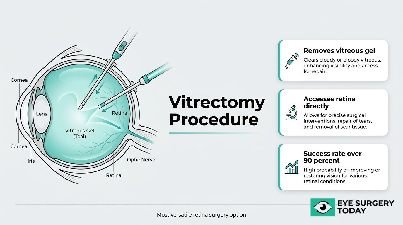
Vitrectomy is a surgical procedure that removes the vitreous gel from the center of the eye to access and repair the retina. Retinal surgeons perform pars plana vitrectomy (PPV) to treat conditions such as macular holes, epiretinal membranes, diabetic vitreous hemorrhage, and retinal detachments. During the procedure, the surgeon replaces the vitreous with a gas bubble, silicone oil, or saline solution to hold the retina in place while it heals. Because this approach provides direct access to the retinal surface, it remains one of the most versatile tools in vitreoretinal surgery. For conditions like macular holes, vitrectomy has demonstrated a success rate of over 90%, according to the American Society of Retina Specialists.
Scleral Buckle Surgery
Scleral buckle surgery is a procedure that repairs retinal detachments by placing a silicone band around the outside of the eye. This band, called a scleral buckle, gently pushes the wall of the eye inward toward the detached retina, closing the retinal break and allowing fluid to reabsorb. The buckle typically remains permanently in place. Surgeons often choose this technique for rhegmatogenous retinal detachments, particularly in younger patients or those without significant vitreous traction. According to a study published in Clinical and Experimental Ophthalmology, scleral buckling has achieved success rates as high as 95%, even at 20-year follow-up. This long track record makes scleral buckling one of the most reliable options for primary retinal detachment repair.
Pneumatic Retinopexy
Pneumatic retinopexy is a minimally invasive, office-based procedure used to repair certain types of retinal detachments. The surgeon injects a gas bubble into the vitreous cavity, which floats upward and presses against the retinal tear, sealing it against the eye wall. Cryopexy or laser is then applied to create a permanent seal around the break. This technique works best for detachments with a single break located in the upper portion of the retina. According to a review in the Indian Journal of Ophthalmology, pneumatic retinopexy achieved a single-operation success rate of 80%, with 98% of cases resolved after reoperations. Its less invasive nature often translates to a shorter recovery period compared to vitrectomy or scleral buckling.
Laser Photocoagulation
Laser photocoagulation is a procedure that uses focused laser energy to seal retinal tears, destroy abnormal blood vessels, or reduce retinal swelling. The laser creates small burns that form scar tissue, which bonds the retina to the underlying tissue and prevents further detachment or leakage. Retinal specialists commonly use this technique for diabetic retinopathy, retinal tears, and retinal vein occlusion. Panretinal photocoagulation (PRP), a specific pattern of widespread laser application, has been a standard treatment for proliferative diabetic retinopathy for decades. When performed early, laser photocoagulation can help stabilize vision and reduce the risk of severe vision loss, though it does not restore vision already lost to retinal damage.
Cryopexy
Cryopexy is a procedure that uses extreme cold to create a scar around a retinal tear or break, sealing the retina to the underlying tissue. A retinal specialist applies a freezing probe, called a cryoprobe, to the outer surface of the eye directly over the tear. The resulting inflammation produces an adhesive scar that prevents fluid from passing through the break and causing detachment. Cryopexy is frequently used alongside pneumatic retinopexy or scleral buckle surgery rather than as a standalone treatment. It may be preferred over laser photocoagulation when media opacity, such as a vitreous hemorrhage, prevents clear visualization of the retina.
Anti-VEGF Intravitreal Injections
Anti-VEGF intravitreal injections are treatments that deliver medication directly into the vitreous cavity to block vascular endothelial growth factor, a protein that drives abnormal blood vessel growth and fluid leakage in the retina. Conditions treated with anti-VEGF therapy include wet age-related macular degeneration, diabetic macular edema, and retinal vein occlusion. According to a study published in Ophthalmology Science, anti-VEGF injections produced more favorable best corrected visual acuity outcomes compared with panretinal photocoagulation at three years for proliferative diabetic retinopathy. Most patients require a series of injections over time, as the therapeutic effect is temporary. Despite not being a surgical procedure in the traditional sense, anti-VEGF therapy has become a cornerstone of modern retinal care.
Understanding each procedure’s mechanism and application helps clarify which approach may be recommended for a specific retinal condition.
Who Is a Good Candidate for Retina Surgery?
A good candidate for retina surgery is someone with a diagnosed retinal condition that threatens vision and whose overall eye health supports surgical intervention. Candidacy depends on factors such as the specific diagnosis, the severity of retinal damage, the patient’s general health, and how quickly treatment begins.
Your retina specialist may recommend surgery if you have a condition such as retinal detachment, macular hole, epiretinal membrane, or advanced diabetic retinopathy that has not responded adequately to less invasive treatments. Patients with sudden symptoms, including new floaters, flashes of light, or a shadow across the visual field, often require urgent evaluation because early intervention can significantly improve outcomes.
Several factors can influence whether surgery is appropriate and what level of visual recovery to expect:
- Macular involvement: When the macula remains attached in retinal detachment cases, the visual prognosis is generally more favorable than when the macula has detached.
- Timing of intervention: Patients who receive prompt treatment before extensive retinal damage occurs tend to achieve better post-operative vision.
- Overall eye health: The presence of other conditions, such as advanced glaucoma or severe corneal disease, may complicate surgical planning and affect candidacy.
- Systemic health considerations: Uncontrolled diabetes, blood clotting disorders, or cardiovascular conditions may require stabilization before retina surgery can proceed safely.
- Patient compliance capacity: Some procedures require strict post-operative positioning, so a patient’s ability to follow recovery protocols factors into the decision.
Not every case carries the same expected outcome. As Dr. John T. Thompson, MD, editor of the ASRS Retina Health Series, notes, “the visual prognosis is more guarded in eyes with massive hemorrhagic choroidal detachments, but most eyes with choroidal detachments have a good visual prognosis.” This underscores why individualized assessment by a retina specialist matters; the same condition can carry very different outlooks depending on severity and complicating factors.
Ultimately, the best way to determine candidacy is through a comprehensive dilated eye examination and diagnostic imaging. Your eye care provider can then weigh the potential benefits of surgery against the risks for your specific situation, helping you make a confident, informed decision about next steps.
What Are the Possible Risks and Complications of Retina Surgery?
The possible risks and complications of retina surgery include both ocular and systemic events, though serious complications remain uncommon. According to a study published in PubMed (PMID 37796532), the overall incidence rate of systemic complications after vitreoretinal surgery was 0.4%, with cardiovascular events accounting for 48.1% of those cases. The following subsections cover procedure-specific risks for vitrectomy, scleral buckle surgery, pneumatic retinopexy, and laser photocoagulation. While retina surgery carries its own unique set of considerations, understanding the specific risks of irrigation-aspiration cataract surgery can help patients differentiate various ophthalmic procedures.
What Risks Are Associated with Vitrectomy?
The risks associated with vitrectomy include cataract formation, retinal detachment, infection, glaucoma, bleeding, and re-opening of a macular hole. Pars plana vitrectomy (PPV) performed for diabetic eye disease, particularly diabetic vitreous hemorrhage, requires results to be measured against national benchmarks to ensure acceptable outcomes. Prolonged face-down positioning after vitrectomy with a gas bubble, sometimes lasting up to two weeks, can also cause discomfort and compliance challenges. Failure to maintain proper positioning may compromise the surgical repair. While most vitrectomy patients recover well, the combination of potential ocular complications and strict postoperative demands makes a thorough discussion with a retina specialist essential before proceeding.
What Risks Are Associated with Scleral Buckle Surgery?
The risks associated with scleral buckle surgery include infection, scleral perforation, extrusion of the buckle element, double vision, refractive changes, and elevated intraocular pressure. Discomfort from the silicone band or sponge placed around the eye can persist in some patients. Despite these potential complications, scleral buckling remains highly effective. According to a study in Clinical & Experimental Ophthalmology, scleral buckling achieves success rates as high as 95% in primary rhegmatogenous retinal detachment repairs, even at 20-year follow-up. The strong long-term track record of this procedure suggests that while risks exist, they are generally manageable when the surgery is performed by an experienced retina surgeon.
What Risks Are Associated with Pneumatic Retinopexy?
The risks associated with pneumatic retinopexy include new retinal breaks, subretinal gas migration, missed breaks, and a higher re-detachment rate compared to other procedures. According to the Indian Journal of Ophthalmology, pneumatic retinopexy had a single-operation success rate of 80%, with 98% of cases ultimately cured after reoperations. The 20% initial failure rate means some patients may need a second procedure, such as vitrectomy or scleral buckling, to achieve full reattachment. Strict head positioning after gas bubble injection is also required, and non-compliance can increase the risk of failure. For appropriately selected detachments, pneumatic retinopexy offers a less invasive option, though patients should understand the possibility of reoperation.
What Risks Are Associated with Laser Photocoagulation?
The risks associated with laser photocoagulation include mild vision loss, reduced peripheral vision, decreased night vision, and inadvertent damage to surrounding retinal tissue. Because the laser creates controlled burns to seal retinal tears or reduce abnormal blood vessel growth, some degree of retinal scarring is expected. In rare cases, choroidal neovascularization or macular edema may develop after treatment. Compared to incisional retina procedures, laser photocoagulation carries a lower overall risk profile since it is non-invasive and typically performed in an outpatient setting. Most patients tolerate the procedure well, though multiple sessions may be necessary for conditions like diabetic retinopathy. Understanding the balance between controlled tissue damage and therapeutic benefit helps set realistic expectations before treatment.
How Should You Prepare for Retina Surgery?
You should prepare for retina surgery by completing a thorough pre-operative evaluation, adjusting medications as directed, and arranging logistics for surgery day and recovery.
Pre-operative evaluation for retina surgery includes a dilated eye exam, optical coherence tomography (OCT) for high-resolution evaluation, and sometimes B-scan ultrasonography, according to EyeWiki (American Academy of Ophthalmology). These imaging tools help your retina specialist map the exact location and severity of the problem before selecting a surgical approach.
Your ophthalmologist may recommend the following preparation steps:
- Disclose all medications and supplements. Blood thinners, aspirin, and certain herbal supplements may need to be paused before surgery to reduce bleeding risk.
- Arrange transportation. You will not be able to drive yourself home after the procedure due to sedation and dilated pupils.
- Follow fasting instructions. Most retina surgeries performed under sedation or general anesthesia require fasting for a set number of hours beforehand.
- Plan for recovery positioning. If vitrectomy with a gas bubble is anticipated, prepare face-down positioning equipment at home in advance.
- Ask about eye drop schedules. Your surgeon may prescribe antibiotic or anti-inflammatory drops to begin before the day of surgery.
Thorough preparation is often underestimated, yet it directly influences both surgical safety and how smoothly recovery begins. Patients who arrive informed and logistically ready tend to experience less anxiety and fewer complications during the immediate post-operative period. With preparation complete, knowing what happens during the procedure itself can further ease any remaining concerns.
What Should You Expect During a Retina Surgery Procedure?
During a retina surgery procedure, you should expect a carefully staged process that includes anesthesia administration, the surgical repair itself, and immediate post-operative monitoring. The specific steps vary depending on the procedure type.
Most retina surgeries are performed as outpatient procedures, meaning patients go home the same day. The retinal surgeon typically uses local anesthesia, sometimes combined with sedation, to keep the eye numb and the patient comfortable throughout. General anesthesia may be used in certain cases, such as for patients who cannot remain still or for particularly complex repairs.
Once anesthesia takes effect, the surgeon accesses the retina through one of several approaches. In a vitrectomy, small incisions are made in the pars plana region of the eye, and microsurgical instruments remove the vitreous gel before repairing the underlying retinal tissue. For scleral buckle surgery, the surgeon works on the exterior of the eye, placing a silicone band around the sclera to indent the eye wall inward toward the detached retina. Pneumatic retinopexy involves injecting a gas bubble directly into the vitreous cavity, which presses the retina back into position. Laser photocoagulation and cryopexy use thermal or freezing energy to seal retinal tears and prevent further detachment.
The duration of retina surgery ranges from approximately 30 minutes for simpler procedures, such as pneumatic retinopexy, to two hours or more for complex vitrectomies involving membrane peeling or extensive retinal repair. Throughout the procedure, the surgeon uses a specialized operating microscope and, in many cases, wide-angle visualization systems for a detailed view of the retinal surface.
After the surgical repair is complete, the eye may be patched or shielded, and the care team monitors the patient briefly before discharge. Patients typically cannot drive themselves home and should arrange transportation in advance. Your retinal surgeon will provide specific post-operative instructions, including any required head positioning if a gas bubble or silicone oil was placed during the procedure.
Understanding what happens in the operating room can help reduce pre-surgical anxiety. With recovery protocols varying by procedure type, knowing the expected timeline helps patients plan ahead.
How Long Does Recovery from Retina Surgery Take?
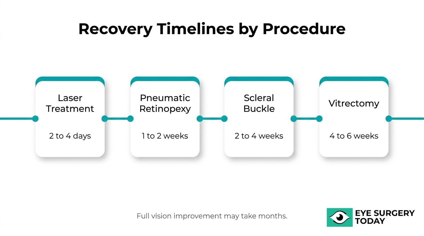
Recovery from retina surgery typically takes 2 to 4 weeks before most patients can return to normal activities, though full vision improvement may take several months. Recovery timelines vary by procedure type.
How Long Is Recovery After Vitrectomy?
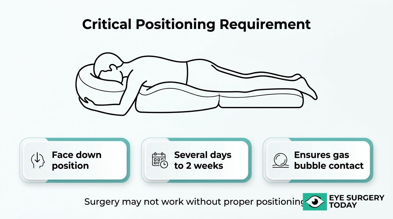
Recovery after vitrectomy generally takes 4 to 6 weeks, making it one of the longer healing periods among retina surgery procedures. Patients who receive a gas bubble during vitrectomy may need to maintain a face-down position for several days, and in some cases up to 2 weeks, to support proper retinal healing. Vision often appears blurry while the gas bubble remains in the eye, gradually clearing as the bubble dissolves. Heavy lifting, strenuous exercise, and air travel are typically restricted during this period. Because vitrectomy involves removing vitreous gel from the eye’s interior, the healing process requires patience as internal structures stabilize. Most patients notice steady visual improvement over the first few months, though the full extent of recovery can extend beyond six months depending on the underlying condition treated.
How Long Is Recovery After Scleral Buckle Surgery?
Recovery after scleral buckle surgery usually takes 2 to 4 weeks for initial healing. According to the American Academy of Ophthalmology, patients who also receive a gas bubble during the procedure may need to maintain strict face-down or sideways positioning to keep the bubble pressing against the retina. Swelling and soreness around the eye are common in the first week, and the eyelids may remain puffy for several days. Return to desk work is often possible within 1 to 2 weeks, while physically demanding jobs may require a longer absence. Vision can fluctuate during the early recovery period as the eye adjusts to the buckle. Complete visual stabilization often takes 3 to 6 months. For many patients, this procedure represents one of the more uncomfortable early recoveries due to external manipulation of the eye wall, though discomfort is manageable with prescribed medication.
How Long Is Recovery After Pneumatic Retinopexy?
Recovery after pneumatic retinopexy is typically shorter than other retinal detachment repairs, with most patients resuming light activities within 1 to 2 weeks. Strict head positioning is required for several days to keep the injected gas bubble pressed against the retinal tear. The bubble gradually absorbs on its own over 2 to 6 weeks. Air travel is not permitted until the gas bubble fully dissolves, as altitude changes can cause dangerous pressure increases inside the eye. Because this procedure is performed in an office setting rather than an operating room, initial recovery tends to involve less postoperative discomfort. Vision improvement occurs progressively as the bubble shrinks and the retina heals against the eye wall.
How Long Is Recovery After Laser Photocoagulation?
Recovery after laser photocoagulation is the shortest among retina surgery procedures, with most patients returning to normal activities within a few days. Vision may appear slightly blurry for 24 to 48 hours after treatment, and mild discomfort or light sensitivity can occur briefly. Because laser photocoagulation is a noninvasive outpatient procedure, no surgical incision needs to heal. According to data published by Ophthalmology Advisor, best-corrected visual acuity improved in all patients after retinal detachment repair, from 1.67±0.97 logMAR before surgery to 0.21±0.22 logMAR at the endpoint more than 6 months later, underscoring the importance of long-term follow-up regardless of the initial procedure. Multiple sessions may be needed for conditions like diabetic retinopathy, with each session requiring minimal downtime.
With recovery timelines understood, ongoing eye wellness and consistent post-operative care help protect these results long-term.
What Does Post-Operative Care Involve After Retina Surgery?
Post-operative care after retina surgery involves strict positioning requirements, prescribed eye drops, activity restrictions, and scheduled follow-up visits to protect the surgical repair during healing.
Unlike cataract surgery, which replaces a cloudy lens, or LASIK, which reshapes the cornea, retina surgery involves internal vitreous removal or external scleral reinforcement to repair the neural tissue of the eye. This distinction makes post-operative compliance especially critical; the delicate retinal tissue requires a controlled healing environment that patients actively maintain through daily behaviors.
According to the American Academy of Ophthalmology, after retinal surgery with a gas bubble, patients must stay face down or sideways at all times, as the surgery may not work if positioning instructions are not followed. This face-down requirement can last several days to two weeks, depending on the procedure and condition treated.
Key components of post-operative care include:
- Positioning compliance: Maintaining the prescribed head position allows the gas bubble to press against the retina and hold it in place during healing.
- Prescribed eye drops: Antibiotic and anti-inflammatory drops help prevent infection and reduce swelling at the surgical site.
- Activity restrictions: Heavy lifting, strenuous exercise, and bending at the waist are typically restricted for several weeks.
- Air travel avoidance: Patients with an intraocular gas bubble cannot fly, as altitude changes cause the bubble to expand and raise intraocular pressure dangerously.
- Follow-up appointments: Regular check-ups allow the retina specialist to monitor reattachment, bubble absorption, and early signs of complications.
Because retinal detachment is a sight-threatening condition with an incidence of approximately 1 in 10,000 (with higher rates in males, myopic eyes, or after prior ocular surgery), diligent post-operative care can mean the difference between a successful repair and a failed one. Patients who understand why each restriction exists tend to follow protocols more consistently, which directly supports better visual outcomes.
In clinical practice, the positioning requirement is often the most challenging aspect for patients. Planning ahead with face-down support equipment and arranging help at home during the first week can significantly reduce discomfort and improve adherence to the recovery protocol.
How Does Retina Surgery Differ from Other Eye Surgeries?
Retina surgery differs from other eye surgeries by targeting the delicate neural tissue at the back of the eye rather than the lens or cornea. Unlike cataract surgery, which replaces a cloudy lens, or LASIK, which reshapes the cornea, retina surgery involves internal vitreous removal or external scleral reinforcement to repair retinal damage. These structural differences affect surgical complexity, recovery protocols, and risk profiles. Other procedures, such as eyelid surgery, also differ significantly in their approach, often focusing on cosmetic or functional improvements of the surrounding eye structures rather than the retina itself.
Cataract surgery is one of the most common elective procedures, yet it can occasionally lead to retinal complications. According to an IRIS Registry study, rhegmatogenous retinal detachment occurs in approximately 1 in 500 cataract surgeries among patients older than 40 within one year of the procedure. This connection highlights why retinal health monitoring remains important even after routine anterior segment surgery.
Recovery expectations also vary significantly. Cataract and LASIK patients often resume normal activities within days, while retina surgery may require weeks of restricted positioning, limited physical activity, and gradual visual improvement over months. The precision required to work on tissue measured in microns, combined with post-operative gas bubble tamponade or silicone oil placement, makes vitreoretinal procedures fundamentally more involved than refractive or lens-based surgeries.
For patients weighing their options, understanding these distinctions can help set realistic expectations about surgical timelines, recovery demands, and visual outcomes.
What Potential Vision Outcomes Can Retina Surgery Provide?
Retina surgery can provide a range of vision outcomes, from significant visual improvement to stabilization of existing sight. Results vary depending on the condition treated, its severity, and how quickly intervention occurs.
Vision recovery after retina surgery is not uniform. Some patients experience meaningful gains in visual acuity, while others may see modest improvement or, in certain cases, further decline. According to EyeWiki’s review of clinical trials in surgical retina, visual outcomes improved by 10 or more letters in 38% of patients, while 22% experienced a reduction of 10 or more letters. These figures underscore that retina surgery can help preserve or restore functional vision for many individuals, though outcomes are never guaranteed.
Several factors may influence the final visual result:
- Pre-operative visual acuity: Eyes with better vision before surgery tend to achieve stronger post-operative outcomes.
- Timing of intervention: Earlier surgical repair, particularly for retinal detachment, is generally associated with more favorable recovery.
- Condition being treated: Macular hole repair, retinal detachment surgery, and diabetic vitrectomy each carry different outcome profiles.
- Macular involvement: Whether the macula was affected before surgery plays a critical role in determining how much central vision can be recovered.
Setting realistic expectations before any procedure is essential. A retina specialist can help patients understand what degree of improvement is achievable based on their specific diagnosis and ocular health. For many, the primary goal of surgery is preventing further vision loss, with meaningful visual gains representing the best possible scenario rather than a certainty.
Understanding what retina surgery can realistically achieve helps patients weigh their options with greater confidence.
How Should You Evaluate Your Options When Considering Eye Surgery?
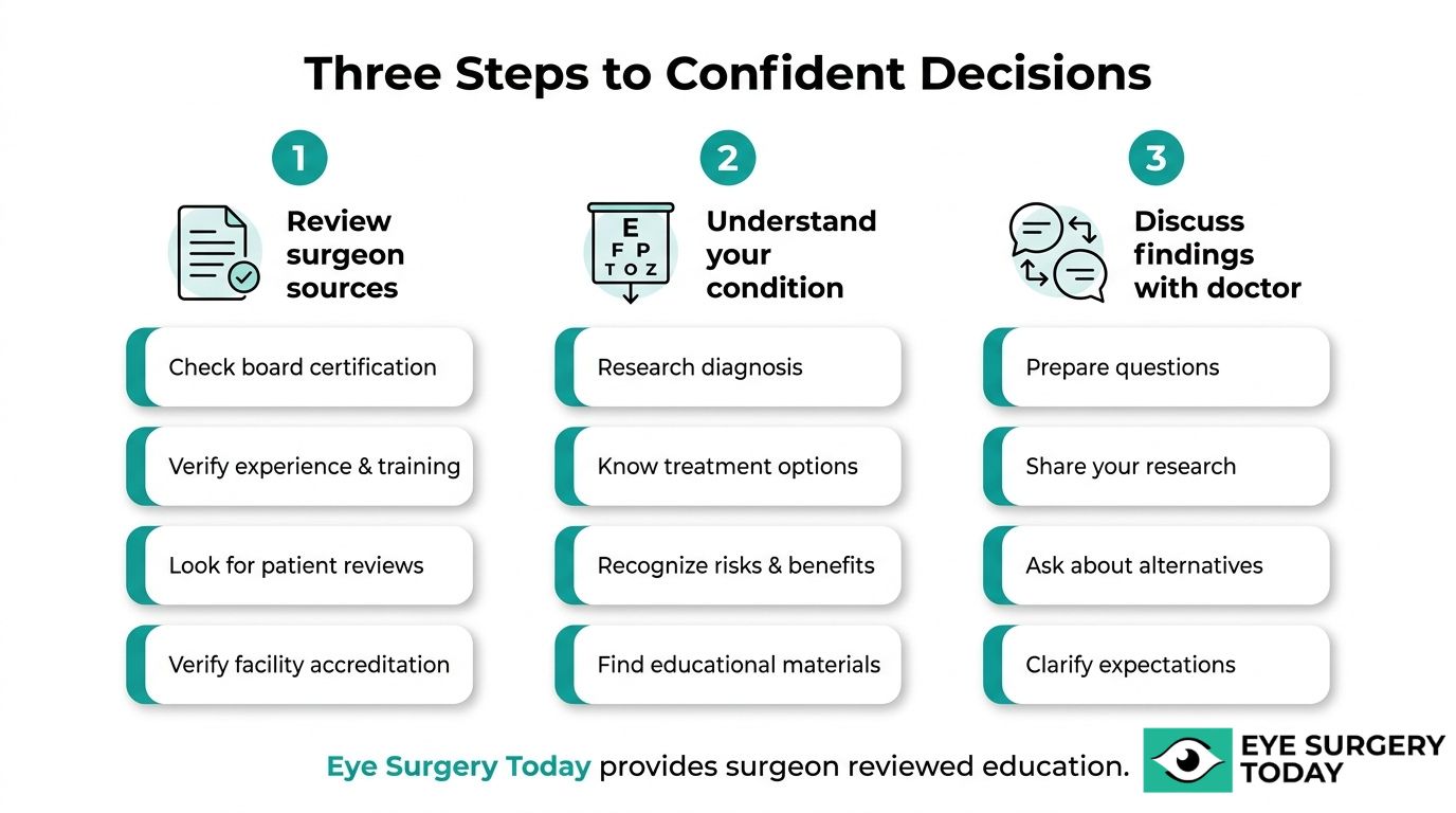
You should evaluate your options when considering eye surgery by reviewing surgeon-reviewed educational resources, understanding your specific condition, and discussing pre-operative findings with your doctor.
Can a Surgeon-Reviewed Education Platform Help You Understand Retina Surgery?
Yes, a surgeon-reviewed education platform can help you understand retina surgery by translating complex clinical information into clear, accessible language. Pre-operative evaluation for retina surgery includes a dilated eye exam, optical coherence tomography (OCT) for high-resolution retinal imaging, and sometimes B-scan ultrasonography, according to EyeWiki, published by the American Academy of Ophthalmology. Platforms that present this type of clinical detail in patient-friendly terms can help individuals grasp what each diagnostic step reveals and why it matters before surgery. Eye Surgery Today provides surgeon-reviewed educational content designed to bridge exactly this knowledge gap, helping patients approach consultations with greater confidence and clarity.
What Are the Key Takeaways About Retina Surgery Procedures, Conditions, and Recovery?
The key takeaways about retina surgery procedures, conditions, and recovery are:
- Retina surgery encompasses several procedures, including vitrectomy, scleral buckle surgery, pneumatic retinopexy, laser photocoagulation, cryopexy, and anti-VEGF intravitreal injections.
- Conditions that may require retina surgery include retinal detachment, macular holes, epiretinal membranes, diabetic retinopathy, retinal vein occlusion, and macular degeneration.
- Recovery timelines vary by procedure, with most patients needing weeks before returning to normal activities and months for full visual improvement.
- Candidacy depends on the specific condition, its severity, and individual health factors that your ophthalmologist will assess during pre-operative evaluation.
- Post-operative care, including strict positioning requirements after gas bubble procedures, directly influences surgical success.
For patients weighing whether retina surgery is right for them, the most practical step is to discuss diagnostic findings and procedure-specific expectations with a retina specialist. Eye Surgery Today offers surgeon-reviewed guides on each of these topics to support informed decision-making.